Skip to navigationSkip to contentSkip to footer
Antibody Arrays
  • Home
  • Multiplex Assays
  • Antibody Arrays

Antibody Arrays

With a huge list of targets, formats, and kits to choose from and no special equipment requirements, you can build a comprehensive view of protein expression levels with a single antibody array study.

Browse Antibody Arrays

Discover More From Less Sample

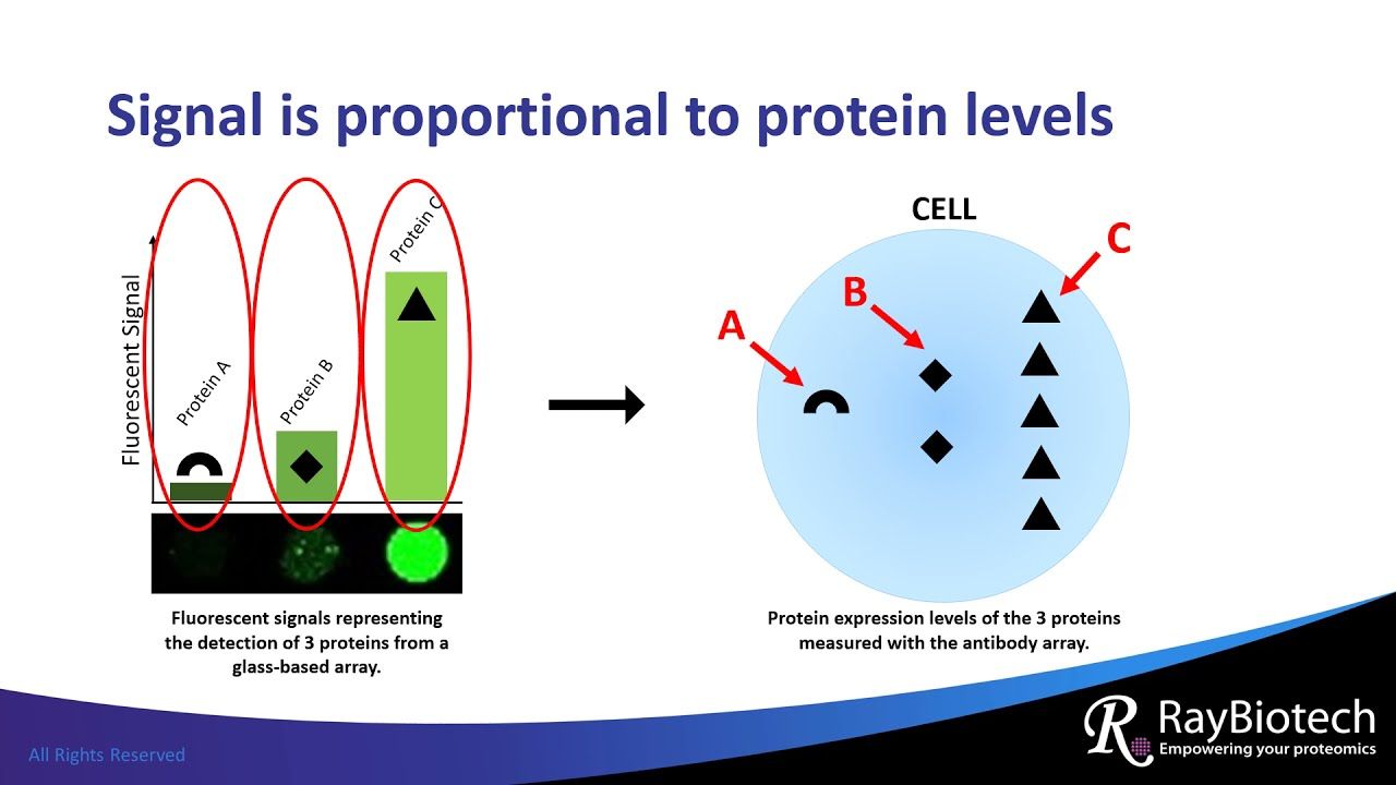
Antibody arrays, also known as antibody microarrays, are multiplex assays that simultaneously measure the expression of multiple proteins, for more comprehensive insight than you can obtain with a single-target ELISA or western blot.

In addition, by measuring expression levels of multiple proteins from a single sample in a single study, you can obtain insight quickly and have more confidence in the comparability of your measurements than when you spend extra time, sample, and effort—and introduce additional variability—normalizing and comparing multiple experiments.

Learn more about multiplex protein detection with antibody arrays

Have Complete Confidence In Your Results

Our arrays are shipped quickly, made in the USA, and produce reproducible results. We want you to succeed and provide support services and data analysis tools from our team of PhD scientists to help you get there. With the same antibody pairs as our ELISAs and validated in the appropriate array format, you know you’re getting reliable products that will set you up for success.

Antibody Array Categories

Cytokine Arrays

Simultaneously measure the levels of multiple cytokines in a range of biological fluids—available in membrane and glass slide formats.

Quantibody® Multiplex ELISA

Achieve quantitative, high-throughput protein expression analysis with an array-based multiplex ELISA system.

RayPlex® Bead Arrays

Using a standard flow cytometer, you can now achieve cytokine detection with unparalleled precision and reproducibility.

Membrane Antibody Arrays

Take advantage of the familiar chemiluminescent western blotting workflow while measuring relative expression of multiple proteins.

Label-based Antibody Arrays

Screen quickly and comprehensively with our largest array—available in membrane and glass slide formats.

Glass Slide Antibody Arrays

Obtain protein expression level data on multiple proteins while conserving precious sample—complementary laser scanning services available.


microarray (1)

FEATURED SERVICE

Array Testing Services

Whether you need extra bandwidth or additional expertise, you can advance confidently with comprehensive Array Testing Services from our experienced scientific team. Simply send us your samples and we’ll use our highly-regarded technology to generate the data you need. We can even handle the analysis through our Biostatistics and Bioinformatics services.

View Array Services

What is an Antibody Array?

Which Antibody Array Fits Your Application?

Applications

Platform

Design Principle

Detection

Data Type

# of Analytes

Quantibody®

Protein Expression Profiling

Slide

Sandwich-based, quadruplicate

Fluorescent

Quantitative

10-1000

L-Series Arrays

Proteome Profiling / Screening

Slide or Membrane

Label-based, duplicate

Fluorescent or Chemiluminescent

Semi-Quantitative

90-8000

C-Series Arrays

Protein Expression Profiling

Membrane

Sandwich-based, duplicate

Chemiluminescent

Semi-Quantitative

10-274

RayPlex® Arrays

Protein Expression Profiling

Microbead

Sandwich-based

Flow Cytometer

Semi-Quantitative

14

Phospho-Arrays

Phosphorylation Profiling

Slide or Membrane

Sandwich-based, quadruplicate

Fluorescent or Chemiluminescent

Semi-Quantitative

17-71

G-Series Arrays

Protein Expression Profiling

Slide

Sandwich-based, duplicate or quadruplicate

Fluorescent

Semi-Quantitative

10-1000

Lectin Arrays

Protein-Lectin Interaction

Slide

Label-based or Sandwich-based

Fluorescent

Semi-Quantitative

70-95

Glycome Arrays

Glycosylation profiling

Slide

Sandwich-based (lectin-ab pair)

Fluorescent

Semi-Quantitative

2000

Protein Arrays

Epitope Mapping; Auto-antibody Profiling / Characterization

Slide

Label-based

Fluorescent

Semi-Quantitative

22-48

How to Choose an Antibody Array

Your Need

Our Solution

I want to screen as many factors as possible (I need a "big net")

L-Series: Label-based Arrays or larger Quantibody® Arrays

I want to focus on a specific pathway or biological process

Pathway-specific arrays (e.g. Inflammation; Apoptosis, etc) or Phosphorylation Arrays

I want to choose a specific panel of markers

Custom Array

I have a limited sample volume

Glass Slide-based Arrays:

  • G-Series
  • L-Series (glass slide)
  • Quantibody® Arrays

I don't have a laser scanner

Membrane-based Arrays:

  • C-Series
  • L-Series (membrane)

Or use our free glass slide scanning service

I want quantitative results

Quantibody® Arrays

I want to identify antibody isotypes

Isotyping Arrays

I want to study protein glycosylation

Glycobiology Arrays (Lectin Arrays, Glycosylation Arrays, Glycan Arrays)

I want to screen protein-protein interactions

Protein Arrays

I have samples from an uncommon species

L-Series: Label-based Arrays or Quantibody® Arrays

I want to do biomarker discovery

Any RayBiotech Array

How Antibody Arrays Can Advance Your Research


FAQ

Frequently Asked Questions

Still have questions?

Contact Us

Quantitative data gives you the actual concentration (pg/ml, for example) of the analyte, while semi-quantitative data gives you the relative fold-change of the analyte compared to the concentration in another sample.

The main difference between the glass-slide and membrane-based arrays is the type of detection used. Glass-slide-based arrays use fluorescence and need a compatible laser scanner, while membrane-based arrays use chemiluminescence, which can be detected with most western blot imaging systems.

Another difference is the required sample size. Glass-slide assays use smaller sample sizes than membrane-based assays. Glass slides require ~70 ul – 100 ul, while the membranes require at least 1 ml, after dilution.

Lastly, glass-slide-based arrays are generally cheaper than membrane-based arrays.

For our membrane-based arrays, any chemiluminescence imaging system such as an X-ray film developer, CCD camera, or gel documentation system should work. Imaging systems using near-infrared flours, such as the Li-Cor Odyssey and Typhoon systems, also work extremely well with our membrane arrays.

For the glass slide-based arrays, you’ll need a gene microarray laser scanner. A list of specifications and compatible laser scanners can be found here:

Compatible Laser Scanners for Glass Slide Arrays

Most of our arrays are compatible with any biological fluid. This includes cell culture media, cell lysates, tissue lysates, and all clarified body fluids (serum, plasma, urine, cerebrospinal fluid, BAL, saliva, tears, etc.)

There are exceptions for label-based arrays (L-Series), which have limited compatibility with lysates and non-serum/plasma body fluids. Our phosphorylation arrays, which detect membrane-bound receptors, are only validated for use with cell and tissue lysates.

In general, any cell-free extract that contains soluble, non-denatured proteins is likely to work with our arrays.

Our tips for sample preparation can be found here:

Tips on Sample Preparation

The positive control spots are standardized amounts of biotinylated IgG. They are used for signal normalization, monitoring of the detection step, and to help orient the array image.

The negative control spots are printed with buffer only, and thus are not expected to give signals. Negative control spots are used for background subtraction.

The areas on the array map labeled as “blank” are empty (there is nothing printed there). Both the negative control and blank spots should give similar intensity values, and either one may be used to represent the background.

For our membrane arrays, data collection can be accomplished by any densitometry software. CCD camera-based imaging systems are typically equipped with a built-in densitometry software application.

Alternatively, there is a free densitometry program available through the NIH (Image J), which can be downloaded here. More information on how to use Image J can be found here.

For glass slide-based arrays, data extraction is accomplished by the software application associated with the laser scanner. For faster spot identification, we can provide you with a free GAL file which can be downloaded here. More information regarding glass slide array data extraction can be found here.

Normalization is used to compare data between arrays (i.e., different samples) by accounting for the differences in signal intensities of the positive control spots on those arrays. We can do this because the positive control is a controlled amount of biotinylated antibody that is attached to the solid support. The amount of signal from each of those spots is dependent on 1) the amount of the reporter (streptavidin-HRP or streptavidin-fluor) bound to that antibody and 2) the length of exposure time (for chemiluminescent detection).

Since these factors proportionally affect the signal intensity of every spot on the array, the differences in the positive control signals between arrays will accurately reflect the differences between other spots on those arrays.

One array must be defined as the "reference array“ to which the signals of other arrays are normalized. It is up to the researcher to define which array should be the reference.

You can calculate the normalized values as follows:
X(nY) = X(Y) x P1/P(Y)

  • P1 = the average signal density of the positive control spots on the reference array
  • P(Y) = the average signal density of the positive control spots on Array Y
  • X(Y) = the signal density for a particular spot on Array for sample “Y”
  • X(nY)= the normalized value for that particular spot "X" on Array for sample “Y"

Please note that RayBiotech’s analysis software tools can perform the normalization automatically. More information about our analysis tools can be found here.

No problem! Simply send your slides to us and we can scan them for you. We will provide you with the scanned image (TIF file) and the densitometry values with full analysis. Learn more about our scanning services here:

Scanning and Data Analysis Services

Different arrays have different sensitivities. You can find more information here:

Quantibody® Array

Human Cytokine Antibody Array

Mouse Cytokine Antibody Array

We’ve created a helpful guide to help you organize your slides. You can find it here:

Quantibody Standard Options

Still have questions?

Contact Us

Related Services

Custom Antibody Arrays

Build an antibody array for the specific targets and formats—membrane, glass slide, beads—your project needs, while leveraging RayBiotech’s validated antibody pairs.

Array Scanning and Analysis

Conserve precious sample while obtaining comprehensive protein expression insights even if you don’t have access to a laser scanner—scanning is free for all RayBio glass slide arrays.

Array Printing Service

We can print an array of any biomolecule, including antibodies, proteins, glycoproteins, glycolipids, complex biological fluids, peptides, or other small molecules.

Related Products

PTM Arrays

Post-Translational Modification (PTM) multiplex assays for acetylation, acylation, nitration, nitrosylation, oxidation, phosphorylation, and SUMOylation.

Peptide Epitope Mapping Arrays

Peptide multiplex assays suitable for antibody epitope mapping and detection of IgG, IgA, IgM, IgE, etc. in serum, plasma, and many other liquid sample types.

ELISA Kits

ELISA Kits that blend quality and performance with competitive pricing and next-day shipping, you’ll generate more reliable data for confident decision-making.

What customers are saying

Quote icon
“It is the only ELISA Array [Mouse Chemokine Array Q1, Cat# QAM-CHE-1] that gives a quantified assessment of 25 chemokines in one go. Protocol is easy to follow and Ray-biotech provides free scanning of slides. It is the best chemokine ELISA Array available.”

Shalini

National Institutes of Health (US)

Quote icon
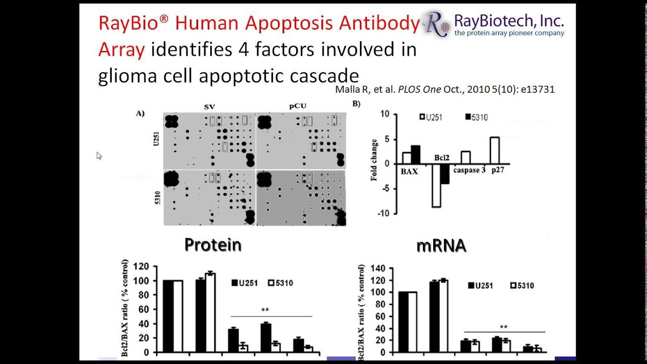
“The human apoptosis array [Human Apoptosis Signaling Array C1, Cat# AAH-APOSIG-1] provides a fast and comprehensive glance about the key signaling involved in the apoptotic pathway. It's a one shot experiment with certain controls already on the blot, pretty simple to execute and normalize. Would recommend it.”

Vinayak

Moffitt Cancer Research Center

Products
  • Multiplex Assays
  • ELISA Kits
  • Proteins and Peptides
  • Antibodies
  • Flow Cytometry
  • Assay Kits
  • Molecular Biology

Sign up to get promotions on your favorite research tools and research updates.

Sign up to Newsletter (opens in new tab)
Services
  • Multiplex Assay Services
  • ELISA Services
  • Antibody Services
  • Custom Protein Services
  • CRO Services
  • Flow Cytometry Services
  • Simoa – Single Molecule Array Services
Resources
  • Manual Protocols
  • Resource Library
©2007-2026 RayBiotech, Inc. All rights reserved.Life Science Web Design By Supreme
  • Privacy Policy
  • Terms & Conditions
  • ISO Certification
  • Risk Free Guarantee
  • Promotions
  • Learning Center
  • Array Analysis Tools
  • Publications / Citations
  • Publications by RayBiotech Scientists
  • About Us
    • About RayBiotech
    • Careers
    • Quality Systems
    • Business Partnerships
    • News & Events
    • Reducing Our Emissions
    Contact Us
    • Contacts
    • Distributors
    • Certified Service Providers
    ISO 13485:2016cGMP
    ISO 17025:2017CLIA
    Need Help?770-729-2992
    MY ACCOUNT

    Your cart is empty.

    RayBiotech
    RayBiotech
    • Products
      • Multiplex Assays
      • ELISA Kits
      • Proteins
      • Antibodies
      • Flow Cytometry
      • Assay Kits
      • Molecular Biology
      • Other Products
      • Human Cytokine Array C5
      • Human IL-6 ELISA
      • Human Inflammation Array Q3
      • Mouse Cytokine Array C3
      • Recombinant SARS-CoV-2 Spike Protein, S1 Subunit
      • Human Phosphorylation Pathway Profiling Array C55
      • Antibody Arrays
      • Proteome Profiling Arrays
      • Cytometric Bead Arrays
      • Epitope Mapping Peptide Arrays
      • Protein Arrays
      • PTM Multiplex Assays
      • NexaTag® Ultrasensitive Multiplex ELISA
      • Sandwich ELISA
      • Competitive ELISA
      • PTM ELISA
      • NexaTag® Ultrasensitive qIPCR ELISA
      • Indirect ELISA
      • Bridging ELISA
      • Cell-Based ELISA
      • Recombinant Proteins
      • GMP Proteins
      • Native Proteins
      • Peptides
      • Primary Antibodies
      • Secondary Antibodies
      • Flow Cytometry Antibodies
      • Isotype Controls
      • Antibody Pairs
      • Flow Cytometry Antibodies
      • Flow Cytometry Reagents
      • Flow Cytometry Assays
      • Cytometric Bead Arrays
      • Fluorescent Beads
      • Quantum Dots
      • Metabolism Assays
      • Ligand Binding Assays
      • Drug Antibody (DA) & ADA Assays
      • Transcription Factor Activity Assays
      • PCR Assays
      • Nucleic Acid Assays
      • Epigenetics
      • Mitochondrial Assays
      • Reagents
      • Cell Culture
      • Biospecimen Samples
      • Sample Collection & Preparation
      • Lysates
      • Mass Spectometry Reagents
      • Magnetic Beads
      • Lab Equipment and Supplies
      • Biological Buffers

      Featured Products

      Human Cytokine Array C5Human IL-6 ELISAHuman Inflammation Array Q3Mouse Cytokine Array C3Recombinant SARS-CoV-2 Spike Protein, S1 SubunitHuman Phosphorylation Pathway Profiling Array C55
    • Services
      • Multiplex Assay Services
      • ELISA Services
      • Protein Services
      • Antibody Services
      • CRO Services
      • Flow Cytometry Services
      • SIMOA – Single Molecule Array
      • Other Services
      • Quantitative Proteomics Services
      • Discovery Proteomics Services
      • Custom Arrays
      • Array Scanning and Analysis
      • Custom Protein Production
      • GMP Protein Production
      • IHC Controls
      • Stable Cell Line Development
      • Molecular Biology Services
      • Antibody Production
      • Recombinant Antibody Expression
      • Bulk Antibody Production
      • Antibody Conjugation Services
      • Cell Biology Services
      • Epitope Mapping Services
      • Biomarker Discovery
      • Cell Biology Services
      • Antibody Drug Development & Characterization
      • Pharmacokinetic and Pharmacodynamic Analysis
      • Assay Development Services
      • Molecular Biology Services
      • Multiomics Services
      • Diagnostic Assay Development
      • GMP Protein Production
      • Biostatistics and Bioinformatics
      • Bulk Antibody Production
      • QC Testing Services
      • Auto-Western Blot Service
      • COVID-19 Pseudovirus Service
      • Biospecimen Samples & Sample Services
      • RNA & DNA Methylation Detection Services
    • Research Areas
      • Cell Signaling Pathways
      • Areas of Interest
      • Akt Signaling Pathway
      • AMPK Signaling pathway
      • ErbB Signaling Pathway
      • Hedgehog Signaling Pathway
      • HIF1-alpha Signaling Pathway
      • IGFR1 Signaling Pathway
      • JAK-STAT Signaling Pathway
      • MAPK Signaling Pathway
      • mTOR Signaling Pathway
      • NF-kappaB Signaling Pathway
      • Notch Signaling Pathway
      • p53 Signaling Pathway
      • PKC Pathway
      • TGF-beta Signaling Pathway
      • Wnt/beta-Catenin Pathway
      • Antibody Drug Development
      • Autoimmunity & Inflammation
      • Cancer
      • Cardiovascular Disease
      • Infectious Disease & Vaccines
      • Neuroscience
      • Obesity
      • Post Translational Modifications
      • RNA & DNA Modifications
    • Resources
      • Assay Picker Tool
      • Sample Shipment Instructions
      • Sample Preparation Tips
      • Publications / Citations
      • Promotions
      • Resource Library
      • Learning Center
      • Manual Protocols
      • Array Analysis Tools
    • Contact Us
      • Distributors & Service Providers
    • About Us
      • Quality Systems
      • Business Partnerships
      • Careers
      • News & Events