RayBiotech

3607 Parkway Ln Ste 200 Norcross, Georgia 30092

www.RayBiotech.com | info@raybiotech.com

770-729-2992 | +1-888-494-8555

ISO 13485:2016 cGMP

Anti-AURKA Antibody

Catalog #: 102-20098

Mouse Anti-AURKA Antibody, 400 µl

Product Description

Specifications

Size400 µL
Estimated Lead Time1-2 weeks
SpeciesHuman
Host SpeciesMouse
Accession Number
O14965
Gene Id
6790
Gene Symbols
AURKA
Protein Name / Synonyms
Aurora kinase A, Aurora 2, Aurora/IPL1-related kinase 1, ARK-1, Aurora-related kinase 1, hARK1, Breast tumor-amplified kinase, Serine/threonine-protein kinase 15, Serine/threonine-protein kinase 6, Serine/threonine-protein kinase aurora-A, AURKA
ClonalityMonoclonal
Clone1364CT291.108.155
IsotypeKappa, IgG1
Recommended ApplicationsWestern Blotting, IHC-P
Shipping TypeBlue ice
Storage-20°C

Description

Introduction

Mitotic serine/threonine kinases that contributes to the regulation of cell cycle progression. Associates with the centrosome and the spindle microtubules during mitosis and plays a critical role in various mitotic events including the establishment of mitotic spindle, centrosome duplication, centrosome separation as well as maturation, chromosomal alignment, spindle assembly checkpoint, and cytokinesis. Required for initial activation of CDK1 at centrosomes. Phosphorylates numerous target proteins, including ARHGEF2, BORA, BRCA1, CDC25B, DLGP5, HDAC6, KIF2A, LATS2, NDEL1, PARD3, PPP1R2, PLK1, RASSF1, TACC3, p53/TP53 and TPX2. Regulates KIF2A tubulin depolymerase activity. Required for normal axon formation. Plays a role in microtubule remodeling during neurite extension. Important for microtubule formation and/or stabilization. Also acts as a key regulatory component of the p53/TP53 pathway, and particularly the checkpoint-response pathways critical for oncogenic transformation of cells, by phosphorylating and stabilizing p53/TP53. Phosphorylates its own inhibitors, the protein phosphatase type 1 (PP1) isoforms, to inhibit their activity. Necessary for proper cilia disassembly prior to mitosis.

Other Information

Antigen Type
Recombinant Protein
Format
Purified monoclonal antibody supplied in PBS with 0.09% (W/V) sodium azide. This antibody is purified through a protein G column, followed by dialysis against PBS.
Calculated Molecular Weight (Da)
45809
Antigen Source
Human

Images

IHC-P (1:25)

Immunohistochemical analysis of paraffin-embedded H. brain section using AURKA Antibody(Cat#102-20098). 102-20098 was diluted at 1:25 dilution. A peroxidase-conjugated goat anti-mouse IgG at 1:400 dilution was used as the secondary antibody, followed by DAB staining.
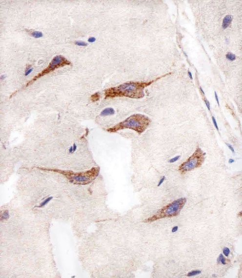
IHC-P

Storage/Stability

2-8°C (short-term); -20°C (long-term)
Expiration:
12 months from the date of shipment when stored properly.