Skip to navigationSkip to contentSkip to footer
Post Translational Modifications
  • Home
  • Applications
  • Post Translational Modifications

Post Translational Modifications

Diversifying our Proteome and Expanding the Functional Capabilities of Proteins

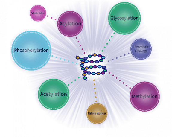
Following synthesis, the majority of proteins will experience modifications. Named for the chronology, these Post-Translational Modifications (PTMs) play a pivotal role in regulating stability, localization, function, and molecular interactions and are responsible for the comparative complexity of the proteome. Over 200 different PTMs have been identified, with the most widely researched being phosphorylation, glycosylation, acetylation, methylation, nitrosylation, acylation, and ubiquitylation. These enzyme-mediated alterations occur at distinct amino acid side chains or peptide linkages and enable the cell to regulate and fine-tune its biological processes. Identifying and understanding these significant mechanisms is critical to the study of basic cell biology as well as in disease pathogenesis.

RayBiotech's suite of PTM profiling tools includes easy-to-use antibody arrays to screen up to 8,000 target proteins for acetylation, palmitoylation, oxidation, glycosylation, or phosphorylation signatures. Additionally, RayBio PTM ELISAs can probe a wide variety of individual signaling events with an efficient, western blot-free workflow. Need extra bandwidth? Any of these assays can be combined into a customized proteomic service to screen and validate protein modifications. To validate individual array targets in your own lab, you can request any single protein modification to be created as a custom, ready-to-use ELISA kit.
View PTM Products Array Services

Featured Publications

See a short list of publications below citing our PTM products.

Importance of glycosylation in the interaction of Tamm-Horsfall protein with collectin-11 and acute kidney injury

Product: Lectin Array 70
Species: Human
Sample type: Urine
View Publication

Palmitoylation of TNF alpha is involved in the regulation of TNF receptor 1 signalling.

Product: Human/Mouse/Rat Phospho-ERK1/2 (T202/Y204) Cell-Based ELISA Kit
Species: Human
Sample type: Conditioned Media (Hela, 3T3L1, ADAM17-deficient (ADAM17zn/zn) and RAW264.7 cell lines)
View Publication

PF4 antagonizes retinal neovascularization via inhibiting PRAS40 phosphorylation in a mouse model of oxygen-induced retinopathy

Product: Human/Mouse AKT Pathway Phosphorylation Array C1
Species: Mouse
Sample type: Vitreous Fluid (retinal fluid)
View Publication

Primary tumor secreted VEGF induces vascular hyperpermeability in premetastatic lung via the occludin phosphorylation/ubiquitination pathway

Product: Mouse SCF ELISA Kit
Species: Mouse
Sample type: Serum, Conditioned Media
View Publication

Inhibiton of RET and JAK2 Signals and Upregulation of VEGFR3 Phosphorylation in Vitro by Galectin-1 in Trophoblast Tumor Cells BeWo

Product: Human RTK Phosphorylation Array C1
Species: Human
Sample type: Cell Lysate (BeWo cell lysate)
View Publication

Deducing the N- and O- glycosylation profile of the spike protein of novel coronavirus SARS-CoV-2

Product: Recombinant SARS-CoV-2 S2 Subunit Protein (full length)
View Publication

Related Resources

Nitric Oxide-mediated PTMs

NO mediates diverse cellular pathways by inducing post-translational modifications (PTMs) in proteins.

Protein PTMs: Translating Diversity to Discovery

These modifications can alter the structure, function and localization of proteins, adding to the spatial and temporal control of protein function.

Related Research Areas

JAK/STAT Signaling Pathway
AMPK Signaling Pathway
TGF-beta Signaling Pathway

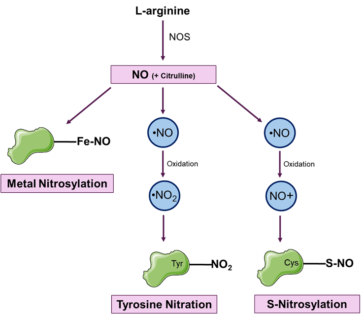
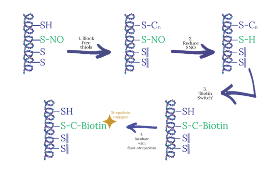
Enzymatic production of nitric oxide (NO) is widespread in cells and tissues. S-nitrosylation is a covalent modification that involves binding a nitric oxide-derived group to the thiol group of cysteine. This process is a vital component of various critical cellular mechanisms, including the regulation of ion channel activity, membrane trafficking, apoptosis, and other post-translational modifications, as well as the maintenance of cellular redox equilibrium. In recent years, it has become clear that dysregulation of S-nitrosylation also contributes to the development of various diseases, including multiple sclerosis, Parkinson's disease, cancer, sickle cell disease, and asthma.

RayBio® Human Protein S-Nitrosylation Antibody Arrays are designed for identifying the relative levels of S-Nitrosylation of cysteine in up to 8,000 different human proteins in various biological samples. The kits use a modified biotin-switch method to label the proteins in the sample. The biotin-labeled samples are then added to antibody arrays, washed, and incubated with a fluorescent dye-conjugated streptavidin for imaging using a laser scanner. The arrays provide a quick and efficient way to monitor changes in protein S-Nitrosylation in experimental model systems.

Browse Nitrosylation Products
Nitric-Oxide PTM Blog Post

Enzymatic production of nitric oxide (NO) is widespread in cells and tissues. S-nitrosylation is a covalent modification that involves binding a nitric oxide-derived group to the thiol group of cysteine. This process is a vital component of various critical cellular mechanisms, including the regulation of ion channel activity, membrane trafficking, apoptosis, and other post-translational modifications, as well as the maintenance of cellular redox equilibrium. In recent years, it has become clear that dysregulation of S-nitrosylation also contributes to the development of various diseases, including multiple sclerosis, Parkinson's disease, cancer, sickle cell disease, and asthma.

RayBio® Human Protein S-Nitrosylation Antibody Arrays are designed for identifying the relative levels of S-Nitrosylation of cysteine in up to 8,000 different human proteins in various biological samples. The kits use a modified biotin-switch method to label the proteins in the sample. The biotin-labeled samples are then added to antibody arrays, washed, and incubated with a fluorescent dye-conjugated streptavidin for imaging using a laser scanner. The arrays provide a quick and efficient way to monitor changes in protein S-Nitrosylation in experimental model systems.

Browse Nitrosylation Products
Nitric-Oxide PTM Blog Post

Phosphorylation is a crucial mechanism in biological systems that regulates many cellular processes involved in growth, apoptosis, and signal transduction. It involves the transfer of a gamma-phosphate to specific amino acids such as serine, threonine, or tyrosine. For a significant number of proteins, phosphorylation is linked with protein activity and is essential to their functionality. Dysregulation of phosphorylation is implicated in a variety of diseases, including cancer, diabetes, and neurological disorders. Abnormal activation or inactivation of specific kinases or phosphatases can result in aberrant signaling pathways, leading to uncontrolled cell growth or impaired cellular function. Therefore, understanding the complex network of phosphorylation events in cells is crucial for developing targeted therapies to treat these diseases.

To aid detection of the phospho-proteome, RayBiotech offers phospho-specific antibody arrays in which up to 8,000 proteins can be screened simultaneously. Alternatively, targeted array panels and phosphorylation ELISAs are available to probe specific signaling pathways related to DNA damage, inflammation, tumorigenesis, and many other processes. Our repertoire of phosphorylation immunoassays features rapid, efficient protocols for high throughput data collection, empowering the researcher with precise, highly sensitive verification of pathway activation, detection of phosphorylation signatures, or enhancement of mass spectrophotometric protein analysis.

Browse Phosphorylation Products

Phosphorylation is a highly reversible state, enabling rapid modulation of cellular activity. The addition and removal of phosphoryl groups to proteins is catalyzed by kinases and phosphatases, respectively.

Glycosylation is a fundamental process of the biosynthetic-secretory pathway of the ER and Golgi apparatus, involving the addition of sugar moieties to specific amino acids. This modification occurs in over half of all expressed proteins and can be categorized into groups based on the amino acid that links to the carbohydrate chain (N-, O-, C-). Each protein has the ability to be glycosylated at multiple sites, potentially with different linkages, enhancing the diversity of the proteome in a way that cannot be achieved by any other post-translational modification. Factors such as enzyme availability, amino acid sequence, and protein conformation all play a role in how glycosylation occurs. Abnormal glycosylation patterns can serve as markers for certain disease states, including cancer metastasis, diabetes mellitus, inflammatory bowel disease, systemic lupus erythematosus, and many other autoimmune conditions.

This diverse structure and complex composition also makes glycoproteins more difficult to analyze, and detection tools for glycobiology research are in limited supply. To address this challenge, RayBiotech has developed three different formats of glycobiology arrays utilizing microarray technology. The Glycan arrays, Glycome Arrays, and Lectin Arrays all utilize glass slide supports with fluorescent detection and serve as powerful tools for researchers to detect glycans and/or glycan-protein interactions.

Browse Glycosylation Products

Glycan ArrayGlycome ArrayLectin Array
Immobilized Molecule
Sugar Moieties (glycans)
Antibodies
Lectins
Species Detected
Not species specific
Human, Mouse, Rat
Not species specific
Spot Replicates
Quadruplicate/Triplicate
Duplicate
Duplicate
Number of Targets
100 or 300
up to 1000
75 or 95

RayBiotech has developed 3 different formats of glycobiology profiling arrays to help researchers detect glycans and/or glycan-protein interactions. All 3 formats feature glass slide supports with fluorescent detection.

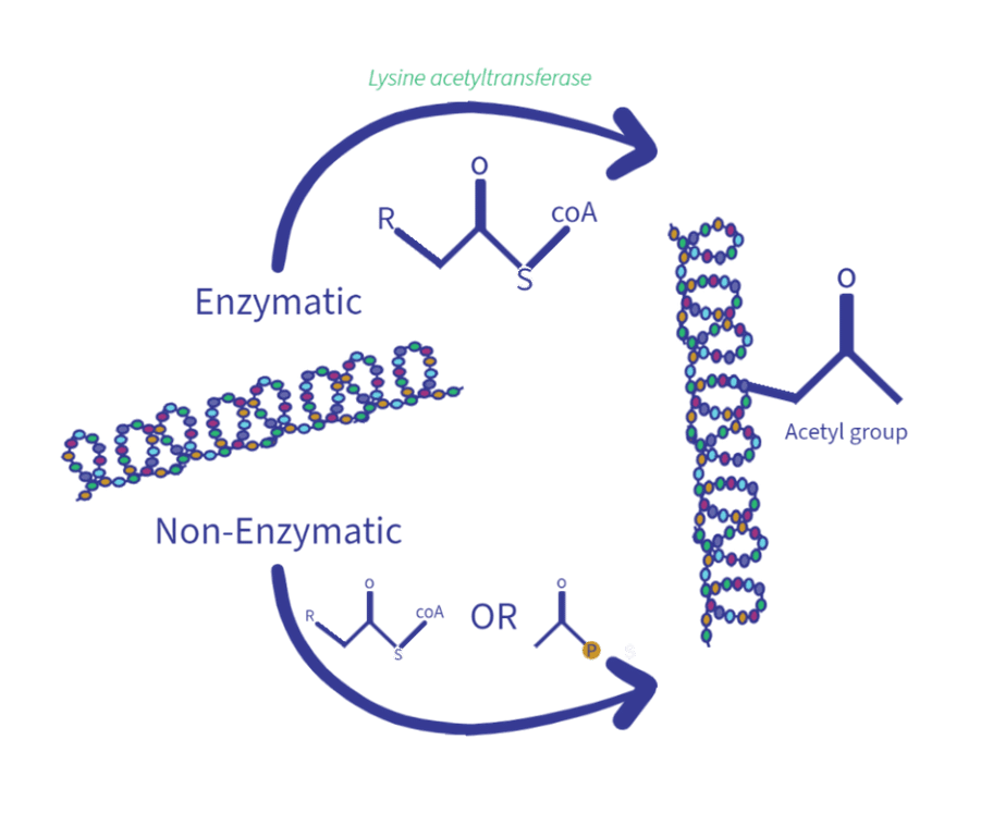
Protein acetylation is a biochemical process whereby a lysine residue on a protein is modified through the addition of an acetyl group. This modification has significant implications for the structure and function of the protein. Initially identified on histones, acetylation increases the size of the side chain, attenuates the amino group's positive charge, and elevates the protein's hydrophobicity. The effects on DNA binding, enzymatic activity, protein-protein interactions, and protein degradation are far-reaching. Acetylation can stimulate gene expression by loosening the chromatin structure and rendering DNA more accessible to transcription factors and other regulatory proteins. Disrupted acetylation has been linked to various diseases, including cancer, neurodegenerative conditions, and metabolic disorders.

RayBio® Human Protein Acetylation Antibody Arrays are designed for identifying the relative levels of acetylation of lysine in up to 8,000 different human proteins in cell lysate. Cell lysate is introduced into the wells of the antibody array glass slide. Then, a biotinylated anti-acetylated-lysine antibody is used to detect acetylated-lysine on target proteins. After incubation with a fluorescent dye-conjugated streptavidin, the slides are imaged using a laser scanner.

Browse Acetylation Products

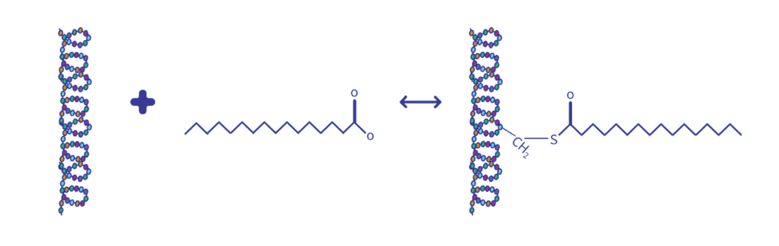
Protein acylation plays a significant role in cell signaling, development, and growth by covalently modifying proteins with lipids. This modification involves the addition of an acyl group, usually a fatty acid, to an amino acid residue. This changes the protein from hydrophilic to hydrophobic at one end, making it easier to interact with membranes. Protein acylation is a crucial aspect of many cellular processes relevant to physiology and diseases such as stability, localization, protein-protein interactions, and protein-DNA interactions. Furthermore, acylation plays a regulatory role in insulin secretion and insulin response pathways. Palmitoylation, the most extensively studied acyl modification, involves the addition of palmitate (a 16-carbon fatty acid) to a cysteine, and occasionally a threonine or serine residue. Unlike other acylation events, protein palmitoylation is reversible. The rapid and dynamic cycling between palmitoylation and de-palmitoylation plays an important role in membrane anchoring and trafficking between intracellular compartments.

RayBio® Human Protein S-Acylation Antibody Arrays are designed for identifying the relative levels of S-acylation of cysteine in up to 8,000 different human proteins in various biological samples. The kits use a modified biotin-switch method to label the samples, wherein the biotin-labeled samples are added to antibody arrays, washed, and incubated with a fluorescent dye-conjugated streptavidin for imaging using a laser scanner. The arrays provide a quick and efficient way to monitor changes in protein S-acylation in experimental model systems.

Browse Acylation Products

Oxidative cell stress arises when the generation of reactive oxygen species (ROSs) and free radicals exceeds the cellular antioxidant defense system's capabilities. Among the key targets of oxidation, proteins take the spotlight due to their abundance and remarkable reactivity towards ROSs. Protein carbonylation occurs when ROSs react with specific amino acid side chains, primarily lysine, arginine, proline, and threonine residues. The presence of carbonylated proteins serves as valuable biomarkers for oxidative stress and has been linked to numerous diseases, including neurodegenerative disorders, cardiovascular diseases, diabetes, and aging-related conditions. Consequently, the detection and quantification of protein carbonylation plays a pivotal role in identifying oxidative stress biomarkers and devising strategies to mitigate their detrimental effects.

RayBio® Human Protein Oxidation Antibody Arrays are designed to assess the relative levels of carbonylation in up to 8,000 different proteins in various biological samples. These innovative kits utilize a modified "biotin probes" method to enable sensitive and specific detection of carbonylated proteins. In this approach, carbonyl groups of proteins react with biotin-hydrazide to form unstable Schiff bases which are subsequently reduced to form stable hydrazine bonds. Following biotin-hydrazide labeling, samples are added to antibody arrays, washed, and incubated with a fluorescent dye-conjugated streptavidin for imaging using a laser scanner.

Browse Oxidation Products

Tyrosine nitration involves the addition of a nitro group (-NO2) to tyrosine residues in proteins, a modification mediated by reactive nitrogen species such as peroxynitrite (ONOO-), nitrogen dioxide (•NO2), and nitronium ion (NO2+). This process occurs under conditions of oxidative and nitrosative stress. While tyrosine is the primary target, other amino acids such as tryptophan, cysteine, and methionine can also undergo nitration due to their susceptibility to RNS. Other molecules susceptible to nitration include lipids, nucleic acids, and small molecules, broadening the impact of nitration in biological systems.

Nitration Arrays
Nitric Oxide-mediated PTM (blog)

Lactylation is a post-translational modification in which a lactyl group, generated from cellular lactate, is covalently attached to lysine residues on proteins. Lactate, once thought of only as a metabolic waste product, is now recognized as a key signaling molecule. Cancer cells often convert glucose to lactate even in the presence of oxygen — a phenomenon known as the Warburg effect — underscoring lactate’s central role in cell metabolism. Through protein lactylation, this abundant metabolite directly modifies proteins and influences their function, linking energy metabolism to biological outcomes. Dysregulated lactylation has been associated with inflammation, immune evasion, fibrosis, and cancer progression, making it an important area of study for disease research.

Protein–Lys–NH₂ + HO–CH(CH₃)–COOH (Lactate) → Protein–Lys–NH–CO–CH(OH)–CH₃ (K-Lac)

RayBio® Human Protein Lactylation Antibody Arrays provide a high-throughput approach to detect lysine lactylation across thousands of human proteins simultaneously. In this assay, cell lysates are incubated on antibody-coated glass slides, and captured proteins are probed with a biotinylated anti-lactyllysine antibody followed by fluorescent streptavidin for scanning. By enabling the detection of lactylation events in up to 8,000 proteins, these arrays offer researchers a convenient way to profile modification patterns and monitor pathway activation in diverse experimental systems.

Browse Lactylation Products
Products
  • Multiplex Assays
  • ELISA Kits
  • Proteins and Peptides
  • Antibodies
  • Flow Cytometry
  • Assay Kits
  • Molecular Biology

Sign up to get promotions on your favorite research tools and research updates.

Sign up to Newsletter (opens in new tab)
Services
  • Multiplex Assay Services
  • ELISA Services
  • Antibody Services
  • Custom Protein Services
  • CRO Services
  • Flow Cytometry Services
  • Simoa – Single Molecule Array Services
Resources
  • Manual Protocols
  • Resource Library
©2007-2026 RayBiotech, Inc. All rights reserved.Life Science Web Design By Supreme
  • Privacy Policy
  • Terms & Conditions
  • ISO Certification
  • Risk Free Guarantee
  • Promotions
  • Learning Center
  • Array Analysis Tools
  • Publications / Citations
  • Publications by RayBiotech Scientists
  • About Us
    • About RayBiotech
    • Careers
    • Quality Systems
    • Business Partnerships
    • News & Events
    • Reducing Our Emissions
    Contact Us
    • Contacts
    • Distributors
    • Certified Service Providers
    ISO 13485:2016cGMP
    ISO 17025:2017CLIA
    Need Help?770-729-2992
    MY ACCOUNT

    Your cart is empty.

    RayBiotech
    RayBiotech
    • Products
      • Multiplex Assays
      • ELISA Kits
      • Proteins
      • Antibodies
      • Flow Cytometry
      • Assay Kits
      • Molecular Biology
      • Other Products
      • Human Cytokine Array C5
      • Human IL-6 ELISA
      • Human Inflammation Array Q3
      • Mouse Cytokine Array C3
      • Recombinant SARS-CoV-2 Spike Protein, S1 Subunit
      • Human Phosphorylation Pathway Profiling Array C55
      • Antibody Arrays
      • Proteome Profiling Arrays
      • Cytometric Bead Arrays
      • Epitope Mapping Peptide Arrays
      • Protein Arrays
      • PTM Multiplex Assays
      • NexaTag® Ultrasensitive Multiplex ELISA
      • Sandwich ELISA
      • Competitive ELISA
      • PTM ELISA
      • NexaTag® Ultrasensitive qIPCR ELISA
      • Indirect ELISA
      • Bridging ELISA
      • Cell-Based ELISA
      • Recombinant Proteins
      • GMP Proteins
      • Native Proteins
      • Peptides
      • Primary Antibodies
      • Secondary Antibodies
      • Flow Cytometry Antibodies
      • Isotype Controls
      • Antibody Pairs
      • Flow Cytometry Antibodies
      • Flow Cytometry Reagents
      • Flow Cytometry Assays
      • Cytometric Bead Arrays
      • Fluorescent Beads
      • Quantum Dots
      • Metabolism Assays
      • Ligand Binding Assays
      • Drug Antibody (DA) & ADA Assays
      • Transcription Factor Activity Assays
      • PCR Assays
      • Nucleic Acid Assays
      • Epigenetics
      • Mitochondrial Assays
      • Reagents
      • Cell Culture
      • Biospecimen Samples
      • Sample Collection & Preparation
      • Lysates
      • Mass Spectometry Reagents
      • Magnetic Beads
      • Lab Equipment and Supplies
      • Biological Buffers

      Featured Products

      Human Cytokine Array C5Human IL-6 ELISAHuman Inflammation Array Q3Mouse Cytokine Array C3Recombinant SARS-CoV-2 Spike Protein, S1 SubunitHuman Phosphorylation Pathway Profiling Array C55
    • Services
      • Multiplex Assay Services
      • ELISA Services
      • Protein Services
      • Antibody Services
      • CRO Services
      • Flow Cytometry Services
      • SIMOA – Single Molecule Array
      • Other Services
      • Quantitative Proteomics Services
      • Discovery Proteomics Services
      • Custom Arrays
      • Array Scanning and Analysis
      • Custom Protein Production
      • GMP Protein Production
      • IHC Controls
      • Stable Cell Line Development
      • Molecular Biology Services
      • Antibody Production
      • Recombinant Antibody Expression
      • Bulk Antibody Production
      • Antibody Conjugation Services
      • Cell Biology Services
      • Epitope Mapping Services
      • Biomarker Discovery
      • Cell Biology Services
      • Antibody Drug Development & Characterization
      • Pharmacokinetic and Pharmacodynamic Analysis
      • Assay Development Services
      • Molecular Biology Services
      • Multiomics Services
      • Diagnostic Assay Development
      • GMP Protein Production
      • Biostatistics and Bioinformatics
      • Bulk Antibody Production
      • QC Testing Services
      • Auto-Western Blot Service
      • COVID-19 Pseudovirus Service
      • Biospecimen Samples & Sample Services
      • RNA & DNA Methylation Detection Services
    • Research Areas
      • Cell Signaling Pathways
      • Areas of Interest
      • Akt Signaling Pathway
      • AMPK Signaling pathway
      • ErbB Signaling Pathway
      • Hedgehog Signaling Pathway
      • HIF1-alpha Signaling Pathway
      • IGFR1 Signaling Pathway
      • JAK-STAT Signaling Pathway
      • MAPK Signaling Pathway
      • mTOR Signaling Pathway
      • NF-kappaB Signaling Pathway
      • Notch Signaling Pathway
      • p53 Signaling Pathway
      • PKC Pathway
      • TGF-beta Signaling Pathway
      • Wnt/beta-Catenin Pathway
      • Antibody Drug Development
      • Autoimmunity & Inflammation
      • Cancer
      • Cardiovascular Disease
      • Infectious Disease & Vaccines
      • Neuroscience
      • Obesity
      • Post Translational Modifications
      • RNA & DNA Modifications
    • Resources
      • Array Picker Tool
      • Sample Shipment Instructions
      • Sample Preparation Tips
      • Publications / Citations
      • Promotions
      • Resource Library
      • Learning Center
      • Manual Protocols
      • Array Analysis Tools
    • Contact Us
      • Distributors & Service Providers
    • About Us
      • Quality Systems
      • Business Partnerships
      • Careers
      • News & Events