CYP1B1 Polyclonal Antibody
Loading...
Loading...
Loading...
Loading...
Anti-CYP1B1 Antibody
| Size | 20 µL, 100 µL |
| Estimated Lead Time | 1-2 weeks |
| Species | Human, Mouse, Rat |
| Host Species | Rabbit |
| Accession Number | Q16678 |
| Gene Id | 1545 |
| Gene Symbols | CYP1B1 |
| Protein Name / Synonyms | CYP1B1, ASGD6, CP1B, CYPIB1, GLC3A, P4501B1, cytochrome P450 1B1 |
| Clonality | Polyclonal |
| Isotype | IgG |
| Purity | Affinity purification |
| Buffers | PBS with 0.02% sodium azide, 50% glycerol, pH7.3 |
| Recommended Applications | Western Blotting, IF |
| Research Area | Angiogenesis |
| Shipping Type | Blue ice |
| Storage | -20°C |
Western blot analysis of extracts of various cell lines, using CYP1B1 antibody (144-01377) at 1:1000 dilution.
Secondary antibody: HRP Goat Anti-Rabbit IgG (H+L) at 1:10000 dilution.
Lysates/proteins: 25ug per lane.
Blocking buffer: 3% nonfat dry milk in TBST.
Detection: ECL Basic Kit.
Exposure time: 5s.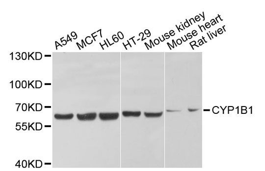
Immunofluorescence analysis of HeLa cells using CYP1B1 antibody (144-01377) at dilution of 1:100 (40x lens). Blue: DAPI for nuclear staining.
Immunofluorescence analysis of NIH/3T3 cells using CYP1B1 antibody (144-01377) at dilution of 1:100 (40x lens). Blue: DAPI for nuclear staining.
Immunofluorescence analysis of PC-12 cells using CYP1B1 antibody (144-01377) at dilution of 1:100 (40x lens). Blue: DAPI for nuclear staining.
Your cart is empty.
Anti-CYP1B1 Antibody