Synonyms
ALS11 antibody, CMT4J antibody, KIAA0274 antibody, SAC3 antibody, dJ249I4.1 antibody, FIG4 antibody, Sac domain-containing inositol phosphatase 3 antibody, SAC domain-containing protein 3 antibody, "FIG4 homolog, SAC1 domain containing lipid phosphatase antibody", "FIG4 homolog, SAC domain containing lipid phosphatase antibody", "phosphatidylinositol 3,5-bisphosphate 5-phosphatase antibody", polyphosphoinositide phosphatase antibody, "FIG4 homolog, SAC1 lipid phosphatase domain containing (S. cerevisiae) antibody"
Specificity
Format
Product Type
Primary antibody
Formulation
0.1M Tris, 0.1M Glycine, 10% Glycerol (pH7). 0.01% Thimerosal was added as a preservative.
Calculated Molecular Weight
104 kDa
Immunogen
Recombinant fragment corresponding to a region within amino acids 135 and 430 of FIG4 (Uniprot ID# Q92562)
Applications
Applications Tested
ICC/IF,IHC (Formalin-fixed paraffin-embedded sections),Western blot
Application Notes
Suggested starting dilutions are as follows: WB: 1:500-1:3000, IHC-P: 1:100-1:1000 [suggested antigen retrieval using heat mediated 10mM Citrate buffer (pH6.0) or Tris-EDTA buffer (pH8.0)], ICC/IF: 1:100-1:1000. Not yet tested in other applications. Optimal working dilutions should be determined experimentally by the end user.
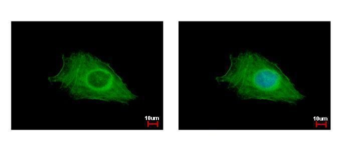
FIG4 antibody detects FIG4 protein at cytoplasm by immunofluorescent analysis.
Sample: HeLa cells were fixed in ice-cold MeOH for 5 min.
Green: FIG4 protein stained by FIG4 antibody (107-10893) diluted at 1:500.
Blue: Hoechst 33342 staining.
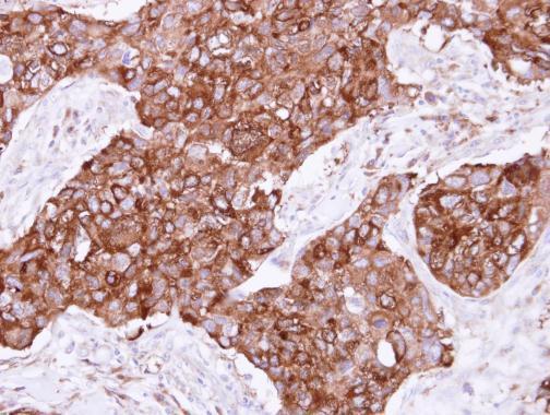
Immunohistochemical analysis of paraffin-embedded Breast ca, using FIG4(107-10893) antibody at 1:500 dilution.
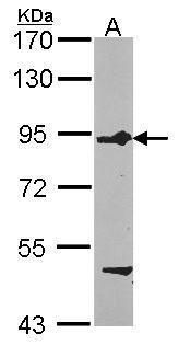
Sample (30 ug of whole cell lysate)
A: HCT116
7.5% SDS PAGE
107-10893 diluted at 1:1000
Storage
Keep as concentrated solution. Aliquot and store at -20°C or below. Avoid multiple freeze-thaw cycles.
Expiration:
12 months from the date of shipment when stored properly.
