RayBiotech

3607 Parkway Ln Ste 200 Norcross, Georgia 30092

www.RayBiotech.com | info@raybiotech.com

770-729-2992 | +1-888-494-8555

ISO 13485:2016 cGMP

Anti-FIP1L1 Antibody

Catalog #: 144-07138

FIP1L1 Polyclonal Antibody

Product Description

Specifications

Size20 µL, 100 µL
Estimated Lead Time1-2 weeks
SpeciesHuman, Mouse, Rat
Host SpeciesRabbit
Accession Number
Q6UN15
Gene Id
81608
Gene Symbols
FIP1L1
Protein Name / Synonyms
FIP1L1, FIP1, Rhe, hFip1, pre-mRNA 3'-end-processing factor FIP1
ClonalityPolyclonal
IsotypeIgG
Purity
Affinity purification
BuffersPBS with 0.02% sodium azide, 50% glycerol, pH7.3
Recommended ApplicationsWestern Blotting, IHC, IF
Shipping TypeBlue ice
Storage-20°C

Description

Background

This gene encodes a subunit of the CPSF (cleavage and polyadenylation specificity factor) complex that polyadenylates the 3' end of mRNA precursors. This gene, the homolog of yeast Fip1 (factor interacting with PAP), binds to U-rich sequences of pre-mRNA and stimulates poly(A) polymerase activity. Its N-terminus contains a PAP-binding site and its C-terminus an RNA-binding domain. An interstitial chromosomal deletion on 4q12 creates an in-frame fusion of human genes FIP1L1 and PDGFRA (platelet-derived growth factor receptor, alpha). The FIP1L1-PDGFRA fusion gene encodes a constitutively activated tyrosine kinase that joins the first 233 amino acids of FIP1L1 to the last 523 amino acids of PDGFRA. This gene fusion and chromosomal deletion is the cause of some forms of idiopathic hypereosinophilic syndrome (HES). This syndrome, recently reclassified as chronic eosinophilic leukemia (CEL), is responsive to treatment with tyrosine kinase inhibitors. Alternative splicing results in multiple transcript variants encoding distinct isoforms.

Dilutions

WB 1:500 - 1:2000
IHC 1:50 - 1:200
IF 1:50 - 1:100

Format

Calculated MW
40kDa/58kDa/65kDa/66kDa
Observed MW
75kDa

Immunogen

Recombinant fusion protein containing a sequence corresponding to amino acids 1-300 of human FIP1L1 (NP_001128409.1).

Modification

Unmodified

Storage

-20°C

Images

Western blot analysis of extracts of various cell lines, using FIP1L1 antibody (144-07138) at 1:1000 dilution.
Secondary antibody: HRP Goat Anti-Rabbit IgG (H+L) at 1:10000 dilution.
Lysates/proteins: 25ug per lane.
Blocking buffer: 3% nonfat dry milk in TBST.
Detection: ECL Basic Kit.
Exposure time: 90s.
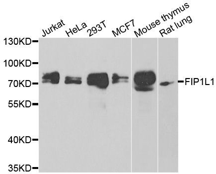
Western blot - FIP1L1 Polyclonal Antibody (144-07138)

Immunohistochemistry of paraffin-embedded mouse liver using FIP1L1 antibody (144-07138) at dilution of 1:100 (40x lens).
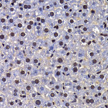
Immunohistochemistry - FIP1L1 Polyclonal Antibody (144-07138)

Immunofluorescence analysis of A549 cells using FIP1L1 antibody (144-07138).
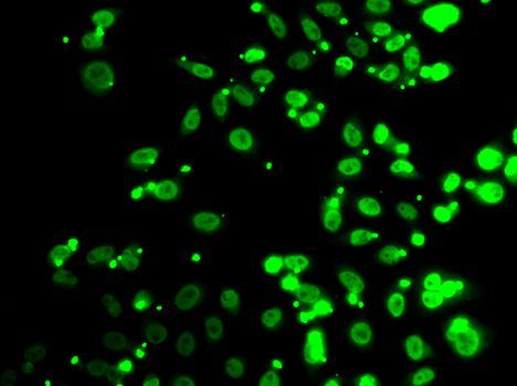
Immunofluorescence - FIP1L1 Polyclonal Antibody (144-07138)

Expiration:
12 months from the date of shipment when stored properly.