3607 Parkway Ln Ste 200 Norcross, Georgia 30092
www.RayBiotech.com | info@raybiotech.com
770-729-2992 | +1-888-494-8555
ISO 13485:2016 cGMP

3607 Parkway Ln Ste 200 Norcross, Georgia 30092
www.RayBiotech.com | info@raybiotech.com
770-729-2992 | +1-888-494-8555
ISO 13485:2016 cGMP
Catalog #: 102-20139
Mouse Anti-HINT1 Antibody, 400 µl
| Size | 400 µL |
| Estimated Lead Time | 1-2 weeks |
| Species | Human, Mouse, Rat |
| Host Species | Mouse |
| Accession Number | P49773 |
| Gene Id | 3094 |
| Gene Symbols | HINT1 |
| Protein Name / Synonyms | Histidine triad nucleotide-binding protein 1, 3---, Adenosine 5'-monophosphoramidase, Protein kinase C inhibitor 1, Protein kinase C-interacting protein 1, PKCI-1, HINT1, HINT, PKCI1, PRKCNH1 |
| Clonality | Monoclonal |
| Clone | 1500CT836.13.93 |
| Isotype | Kappa, IgG1 |
| Recommended Applications | IF, IHC-P, FC, Western Blotting |
| Shipping Type | Blue ice |
| Storage | -20°C |
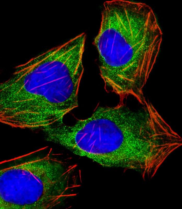
Immunofluorescent analysis of 4% paraformaldehyde-fixed, 0.1% Triton X-100 permeabilized U-2 OS (human bone osteosarcoma cell line) cells labeling HINT1 with 102-20139 at 1/25 dilution, followed by Dylight® 488-conjugated goat anti-mouse IgG (NA166821) secondary antibody at 1/200 dilution (green). Immunofluorescence image showing cytoplasm staining on U-2 OS cell line. Cytoplasmic actin is detected with Dylight® 554 Phalloidin (PD18466410) at 1/100 dilution (red).The nuclear counter stain is DAPI (blue).
Your cart is empty.