RayBiotech

3607 Parkway Ln Ste 200 Norcross, Georgia 30092

www.RayBiotech.com | info@raybiotech.com

770-729-2992 | +1-888-494-8555

ISO 13485:2016 cGMP

Anti-Human CD3 Antibody, Biotinylated

Catalog #: 136-01012

Biotinylated Human CD3 Antibody [clone: OKT3], monoclonal Mouse IgG2a kappa. Validated for flow cytometry.

Product Description

Specifications

Size100 µg, 500 µg
Estimated Lead Time1-2 business days
SpeciesHuman
Host SpeciesMouse
Gene Symbols
CD3
ClonalityMonoclonal
CloneOKT3
ConjugationBiotin
PreparationThe antibody was purified by affinity chromatography, and conjugated with biotin
IsotypeIgG2a Kappa
Format
Liquid
Concentration (lot specific)
0.5 mg/ml
BuffersPBS, pH 7.2-7.4, 0.2% BSA, 0.1% sodium azide
Recommended ApplicationsFlow Cytometry
Research AreaCardiovascular Disease
Shipping TypeBlue ice
Storage4°C

Description

Product Features

  • Serum-free media used for cell culture of hybridomas to minimize bovine antibody contamination
  • Gentle elution at pH 4.0 to minimize antibody degradation
  • Protein A, G & L affinity purification
  • High purity

Typical Data

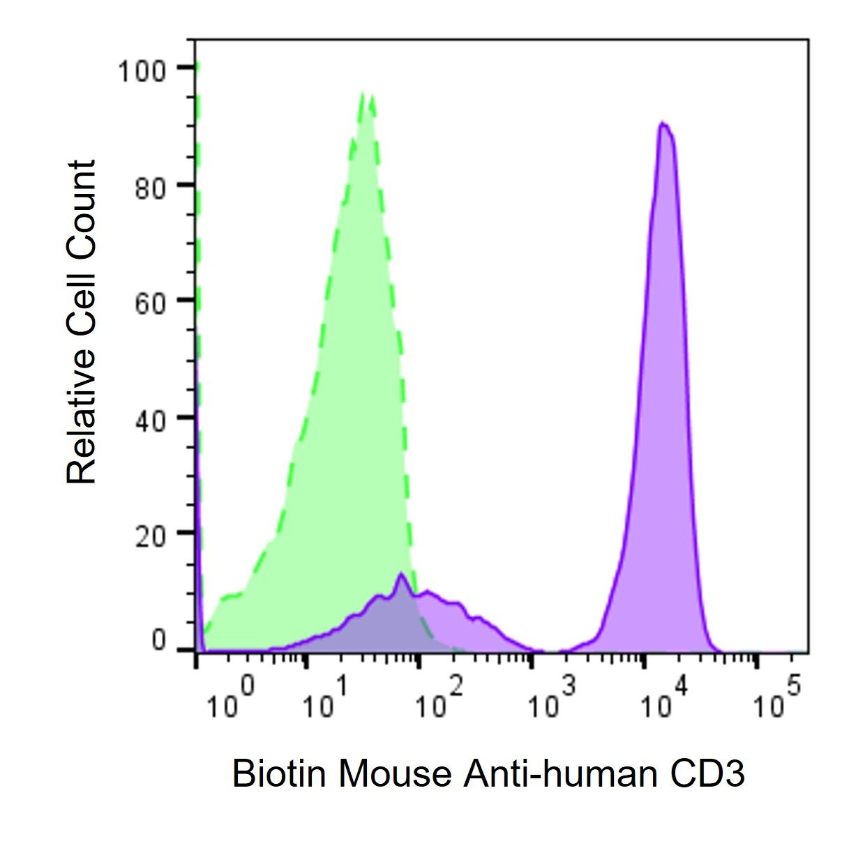
Human PBMCs were stained with either Biotin Mouse Anti-Human CD3 antibody (#136-01012, solid histogram), or Purified Mouse Anti-Human CD3 antibody (#136-00012, dashed histogram), followed by PE-streptavidin.

Description

CD3ε, a subunit of CD3, is a 20 kDa transmembrane protein primarily expressed on pre-mature thymocytes, peripheral mature T cells, and NK-T cells. The CD3 molecules consist of CD3ε, δ, γ, and ζ chains and form a TCR complex by associating with TCR α/β or γ/δ chains. CD3 plays a critical role in TCR signal transduction, T cell activation, and antigen recognition by binding the peptide/MHC antigen complex. The TCR on T cell surface is non-covalently associated with CD3 subunits CD3εγ and CD3εδ heterodimers and the CD3ζζ homodimer, which collectively form the TCR-CD3 complex. Antigen presentation of peptide-loaded MHC molecules by APCs, along with other co-stimulation signals, leads to activation of T cells. This antibody can recognize human CD3ε. It can also activate T cells in vitro together with anti-human CD28.

Recommended Usage

Each lot of antibody has been tested by flow cytometry staining. We suggest use ≤ 0.5 µg per million cells in 100 µl volume of staining buffer. Researchers are recommended to further titrate this antibody at the optimal concentration for each specific application.

Storage / Stability

Stored at 2 °C - 8 °C, protected from prolonged light exposure. DO NOT FREEZE.