RayBiotech

3607 Parkway Ln Ste 200 Norcross, Georgia 30092

www.RayBiotech.com | info@raybiotech.com

770-729-2992 | +1-888-494-8555

ISO 13485:2016 cGMP

Anti-Human CD44 Antibody, Purified

Catalog #: 136-00016

Human CD44 Antibody [clone: FW11-10-03], monoclonal Mouse IgG2a kappa. Validated for flow cytometry.

Product Description

Specifications

Size100 µg, 500 µg
Estimated Lead Time1-2 business days
SpeciesHuman
Host SpeciesMouse
Gene Symbols
CD44
ClonalityMonoclonal
CloneFW11-10-03
PreparationThe antibody was purified by affinity chromatography.
IsotypeIgG2a Kappa
Format
Lyophilized
Concentration (lot specific)
Lyophilized
BuffersPBS, pH 7.2-7.4; Carrier-free
Recommended ApplicationsFlow Cytometry
Reconstitution
Reconstitute in desired amount of PBS.
Research AreaCardiovascular Disease
Shipping TypeAmbient temperature
Storage-20°C

Description

Product Features

  • Serum-free media used for cell culture of hybridomas to minimize bovine antibody contamination
  • Gentle elution at pH 4.0 to minimize antibody degradation
  • Protein A, G & L affinity purification
  • High purity
  • Carrier free
  • Endotoxin-free

Typical Data

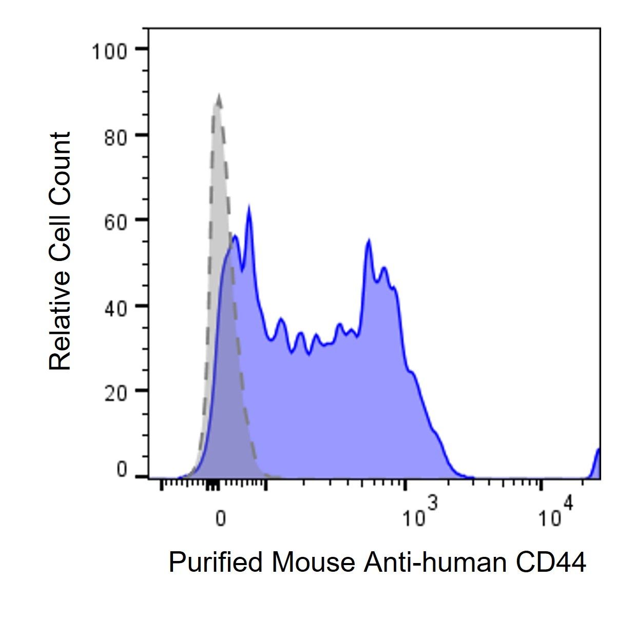
PHA stimulated (3 days) human PBMCs were stained with either Purified Mouse Anti-Human CD44 primary antibody (#136-00016, blue histogram), or mouse IgG isotype control (gray histogram), followed by anti-mouse AF647 IgG/IgM secondary antibody.

Description

CD44 is an 80-95 kDa glycoprotein expressed by a variety of hematopoietic and non-hematopoietic cells including all leukocytes, endothelial cells, hepatocytes, and mesenchymal cells. CD44 combined with CD62L, also called L-selectin, has been used as a T and B cell activation marker. CD44 expression is upregulated in naive T cells upon activation, and high CD44 expression is maintained on memory T cells. Therefore, CD44 is often used to discriminate CD44hi antigen‚¬€œexperienced T cells from CD44low (CD62Lhi) naive T cells. CD44 is also an adhesion molecule involved in leukocyte attachment to endothelial cells, homing to peripheral lymphoid organs and sites of inflammation, and aggregating leukocytes.

Recommended Usage

Each lot of antibody has been tested by flow cytometry staining. We suggest use ≤ 0.5 µg per million cells in 100 µl volume of staining buffer. Researchers are recommended to further titrate this antibody at the optimal concentration for each specific application.

Storage / Stability

Keep the lyophilized antibodies at -20 °C. Once reconstituted with PBS, store at 2 °C - 8 °C for less than 3 weeks or freeze at -20°C for long term storage. Please limit freeze and thaw cycles.