3607 Parkway Ln Ste 200 Norcross, Georgia 30092
www.RayBiotech.com | info@raybiotech.com
770-729-2992 | +1-888-494-8555
ISO 13485:2016 cGMP

3607 Parkway Ln Ste 200 Norcross, Georgia 30092
www.RayBiotech.com | info@raybiotech.com
770-729-2992 | +1-888-494-8555
ISO 13485:2016 cGMP
Catalog #: 136-00047
Human CD56 Antibody [clone: 5.1H11], monoclonal Mouse IgG1 kappa. Validated for flow cytometry.
| Size | 100 µg, 500 µg |
| Estimated Lead Time | 1-2 business days |
| Species | Human |
| Host Species | Mouse |
| Protein Name / Synonyms | CD56, NCAM |
| Clonality | Monoclonal |
| Clone | 5.1H11 |
| Preparation | The antibody was purified by affinity chromatography. |
| Isotype | IgG1 Kappa |
| Format | Lyophilized |
| Concentration (lot specific) | Lyophilized |
| Buffers | PBS, pH 7.2-7.4; Carrier-free |
| Recommended Applications | Flow Cytometry |
| Reconstitution | Reconstitute in desired amount of PBS. |
| Research Area | Cardiovascular Disease |
| Shipping Type | Ambient temperature |
| Storage | -20°C |
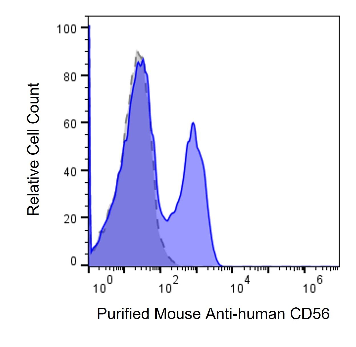
Human PBMCs were stained with either Purified Mouse Anti-Human CD56 primary antibody (#136-00047, blue histogram), or mouse IgG isotype control (gray histogram), followed by AF 647 anti-mouse IgG/IgM secondary antibody.
Your cart is empty.