RayBiotech

3607 Parkway Ln Ste 200 Norcross, Georgia 30092

www.RayBiotech.com | info@raybiotech.com

770-729-2992 | +1-888-494-8555

ISO 13485:2016 cGMP

Anti-KSR1 (N-term A7) Antibody

Catalog #: 102-27397

Rabbit Anti-KSR1 (N-term A7) Antibody, 400 µl

Product Description

Specifications

Size400 µL
Estimated Lead Time1-2 weeks
SpeciesHuman
Host SpeciesRabbit
Accession Number
Q8IVT5
Gene Id
8844
Gene Symbols
KSR1
Protein Name / Synonyms
Kinase suppressor of Ras 1, KSR1, KSR
ClonalityPolyclonal
CloneRB3309
IsotypeKappa, Rabbit
Recommended ApplicationsWestern Blotting, IHC-P
Shipping TypeBlue ice
Storage-20°C

Description

Introduction

Protein kinases are enzymes that transfer a phosphate group from a phosphate donor, generally the g phosphate of ATP, onto an acceptor amino acid in a substrate protein. By this basic mechanism, protein kinases mediate most of the signal transduction in eukaryotic cells, regulating cellular metabolism, transcription, cell cycle progression, cytoskeletal rearrangement and cell movement, apoptosis, and differentiation. With more than 500 gene products, the protein kinase family is one of the largest families of proteins in eukaryotes. The family has been classified in 8 major groups based on sequence comparison of their tyrosine (PTK) or serine/threonine (STK) kinase catalytic domains. The STE group (homologs of yeast Sterile 7, 11, 20 kinases) consists of 50 kinases related to the mitogen-activated protein kinase (MAPK) cascade families (Ste7/MAP2K, Ste11/MAP3K, and Ste20/MAP4K). MAP kinase cascades, consisting of a MAPK and one or more upstream regulatory kinases (MAPKKs) have been best characterized in the yeast pheromone response pathway. Pheromones bind to Ste cell surface receptors and activate yeast MAPK pathway.

Other Information

NCBI Accession #
NP_055053.1
Antigen Type
Synthetic Peptide
Format
Purified polyclonal antibody supplied in PBS with 0.09% (W/V) sodium azide. This antibody is purified through a protein A column, followed by peptide affinity purification.
Calculated Molecular Weight (Da)
102160
Antigen Source
Human

Images

IHC-P (1:50~100)

Formalin-fixed and paraffin-embedded human cancer tissue reacted with the primary antibody, which was peroxidase-conjugated to the secondary antibody, followed by DAB staining. This data demonstrates the use of this antibody for immunohistochemistry; clinical relevance has not been evaluated. BC = breast carcinoma; HC = hepatocarcinoma.
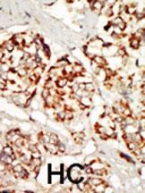
IHC-P

Storage/Stability

2-8°C (short-term); -20°C (long-term)
Expiration:
12 months from the date of shipment when stored properly.