3607 Parkway Ln Ste 200 Norcross, Georgia 30092
www.RayBiotech.com | info@raybiotech.com
770-729-2992 | +1-888-494-8555
ISO 13485:2016 cGMP

3607 Parkway Ln Ste 200 Norcross, Georgia 30092
www.RayBiotech.com | info@raybiotech.com
770-729-2992 | +1-888-494-8555
ISO 13485:2016 cGMP
Catalog #: 102-23002
Rabbit Anti-LC3 (APG8A) (P45) Antibody, 400 µl
| Size | 400 µL |
| Estimated Lead Time | 1-2 weeks |
| Species | Human |
| Host Species | Rabbit |
| Accession Number | Q9H492|Q9GZQ8|A6NCE7|Q9H492;Q9GZQ8|Q62625|Q9CQV6|O41515|Q6XVN8|Q91VR7|Q2HJ23|Q6NX90 |
| Protein Name / Synonyms | Microtubule-associated proteins 1A/1B light chain 3A, Autophagy-related protein LC3 A, Autophagy-related ubiquitin-like modifier LC3 A, MAP1 light chain 3-like protein 1, MAP1A/MAP1B light chain 3 A, MAP1A/MAP1B LC3 A, Microtubule-associated protein 1 light chain 3 alpha, MAP1LC3A |
| Clonality | Polyclonal |
| Clone | RB11840 |
| Isotype | Kappa, Rabbit |
| Recommended Applications | Western Blotting, IHC-P, IF |
| Research Area | Autophagy |
| Shipping Type | Blue ice |
| Storage | -20°C |
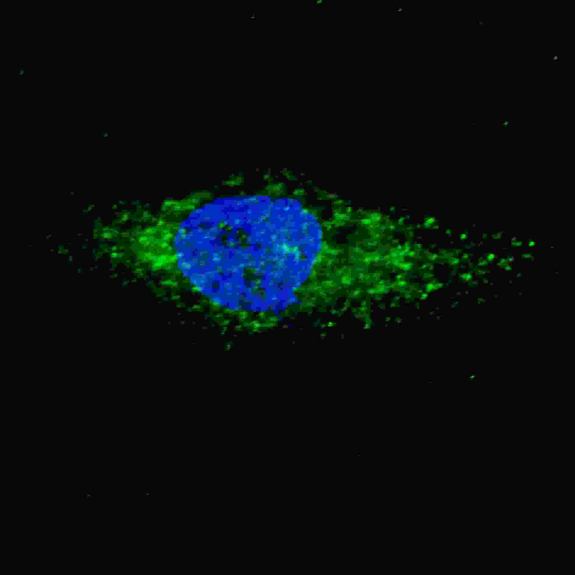
Fluorescent image of U251 cells stained with 102-23002 LC3 (APG8A) (P45) antibody. U251 cells were treated with Chloroquine (50 ?M,16h), then fixed with 4% PFA (20 min), permeabilized with Triton X-100 (0.2%, 30 min). Cells were then incubated with 102-23002 LC3 (APG8A) (P45) primary antibody (1:100, 2 h at room temperature). For secondary antibody, Alexa Fluor® 488 conjugated donkey anti-rabbit antibody (green) was used (1:1000, 1h). Nuclei were counterstained with Hoechst 33342 (blue) (10 ?g/ml, 5 min). LC3 immunoreactivity is localized to autophagic vacuoles in the cytoplasm of U251 cells.
Your cart is empty.