Anti-LC3 (APG8C) (N-term) Antibody
Catalog #: 102-13641
Rabbit Anti-LC3 (APG8C) (N-term) Antibody, 400 ul
Product Description
Specifications
| Size | 400 µL |
| Estimated Lead Time | 1-2 weeks |
| Species | Human, Mouse, Rat |
| Host Species | Rabbit |
| Accession Number | Q9BXW4 |
| Gene Id | 440738 |
| Gene Symbols | MAP1LC3C |
| Protein Name / Synonyms | LC3 (APG8C) (N-term) |
| Clonality | Polyclonal |
| Preparation | This LC3 antibody is generated from rabbits immunized with a KLH conjugated synthetic peptide between 1-30 amino acids from the N-terminal region of human LC3. |
| Concentration (lot specific) | 2.0 mg/ml |
| Recommended Applications | Western Blotting, IHC-P |
| Research Area | Autophagy |
| Shipping Type | Blue ice |
| Storage | -20°C |
Description
Introduction
Macroautophagy is the major inducible pathway for the general turnover of cytoplasmic constituents in eukaryotic cells, it is also responsible for the degradation of active cytoplasmic enzymes and organelles during nutrient starvation. Macroautophagy involves the formation of double-membrane bound autophagosomes which enclose the cytoplasmic constituent targeted for degradation in a membrane bound structure, which then fuse with the lysosome (or vacuole) releasing a single-membrane bound autophagic bodies which are then degraded within the lysosome (or vacuole). MAP1A and MAP1B are microtubule-associated proteins which mediate the physical interactions between microtubules and components of the cytoskeleton. These proteins are involved in formation of autophagosomal vacuoles (autophagosomes). MAP1A and MAP1B each consist of a heavy chain subunit and multiple light chain subunits. MAP1LC3c is one of the light chain subunits and can associate with either MAP1A or MAP1B. The precursor molecule is cleaved by APG4B/ATG4B to form the cytosolic form, LC3-I. This is activated by APG7L/ATG7, transferred to ATG3 and conjugated to phospholipid to form the membrane-bound form, LC3-II.
Specificity
Other Names
MAP1LC3C; Microtubule-associated proteins 1A/1B light chain 3C; Autophagy-related protein LC3 C; Autophagy-related ubiquitin-like modifier LC3 C; MAP1 light chain 3-like protein 3; MAP1A/MAP1B light chain 3 C; Microtubule-associated protein 1 light chain 3 gamma
NCBI Accession #
NP_001004343.1
Format
Type
Purified Rabbit Polyclonal Antibody (Pab)
Calculated Molecular Weight (Da)
16852
Recommended Dilutions
WB: 1:1000; IHC: 1:50-100
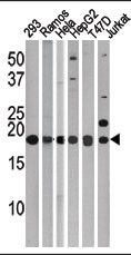 | The anti-LC3 (APG8c) Pab (Cat# 102-13641) is used in Western blot to detect LC3 (APG8c) in, from left to right, 293, Ramos, Hela, HepG2, T47d, and Jurkat tissue lysates. LC3 (APG8c)(arrow) was detected using the purified Pab. |
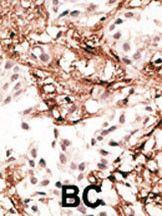 | Formalin-fixed and paraffin-embedded human cancer tissue reacted with the primary antibody, which was peroxidase-conjugated to the secondary antibody, followed by AEC staining. This data demonstrates the use of this antibody for immunohistochemistry; clinical relevance has not been evaluated. BC = breast carcinoma; HC = hepatocarcinoma. |
Antigen Source
HUMAN
Storage/Stability
2-8°C (short-term); -20°C (long-term)
Expiration:
12 months from the date of shipment when stored properly.
