RayBiotech

3607 Parkway Ln Ste 200 Norcross, Georgia 30092

www.RayBiotech.com | info@raybiotech.com

770-729-2992 | +1-888-494-8555

ISO 13485:2016 cGMP

Anti-MAPK3/1 (Center) Antibody

Catalog #: 102-20274

Mouse Anti-MAPK3/1 (Center) Antibody, 200 µl

Product Description

Specifications

Size200 µL
Estimated Lead Time1-2 weeks
SpeciesHuman, Mouse
Host SpeciesMouse
Accession Number
P27361|P28482|Q63844|P63086|P40417|P46196|P63085|P26696|P21708
Gene Id
5595
Gene Symbols
MAPK3
Protein Name / Synonyms
Mitogen-activated protein kinase 3, MAP kinase 3, MAPK 3, 2.7.11.24, ERT2, Extracellular signal-regulated kinase 1, ERK-1, Insulin-stimulated MAP2 kinase, MAP kinase isoform p44, p44-MAPK, Microtubule-associated protein 2 kinase, p44-ERK1, MAPK3, ERK1, PRKM3
ClonalityMonoclonal
Clone1129CT3.4.3
IsotypeKappa, IgG1
Recommended ApplicationsIF, IHC-P, Western Blotting
Research AreaMAPK Signaling
Shipping TypeBlue ice
Storage-20°C

Description

Introduction

Serine/threonine kinase which acts as an essential component of the MAP kinase signal transduction pathway. MAPK1/ERK2 and MAPK3/ERK1 are the 2 MAPKs which play an important role in the MAPK/ERK cascade. They participate also in a signaling cascade initiated by activated KIT and KITLG/SCF. Depending on the cellular context, the MAPK/ERK cascade mediates diverse biological functions such as cell growth, adhesion, survival and differentiation through the regulation of transcription, translation, cytoskeletal rearrangements. The MAPK/ERK cascade plays also a role in initiation and regulation of meiosis, mitosis, and postmitotic functions in differentiated cells by phosphorylating a number of transcription factors. About 160 substrates have already been discovered for ERKs. Many of these substrates are localized in the nucleus, and seem to participate in the regulation of transcription upon stimulation. However, other substrates are found in the cytosol as well as in other cellular organelles, and those are responsible for processes such as translation, mitosis and apoptosis. Moreover, the MAPK/ERK cascade is also involved in the regulation of the endosomal dynamics, including lysosome processing and endosome cycling through the perinuclear recycling compartment (PNRC); as well as in the fragmentation of the Golgi apparatus during mitosis. The substrates include transcription factors (such as ATF2, BCL6, ELK1, ERF, FOS, HSF4 or SPZ1), cytoskeletal elements (such as CANX, CTTN, GJA1, MAP2, MAPT, PXN, SORBS3 or STMN1), regulators of apoptosis (such as BAD, BTG2, CASP9, DAPK1, IER3, MCL1 or PPARG), regulators of translation (such as EIF4EBP1) and a variety of other signaling-related molecules (like ARHGEF2, FRS2 or GRB10). Protein kinases (such as RAF1, RPS6KA1/RSK1, RPS6KA3/RSK2, RPS6KA2/RSK3, RPS6KA6/RSK4, SYK, MKNK1/MNK1, MKNK2/MNK2, RPS6KA5/MSK1, RPS6KA4/MSK2, MAPKAPK3 or MAPKAPK5) and phosphatases (such as DUSP1, DUSP4, DUSP6 or DUSP16) are other substrates which enable the propagation the MAPK/ERK signal to additional cytosolic and nuclear targets, thereby extending the specificity of the cascade.

Other Information

Antigen Type
Synthesized Peptide
Format
Purified monoclonal antibody supplied in PBS with 0.09% (W/V) sodium azide. This antibody is purified through a protein G column, followed by dialysis against PBS.
Calculated Molecular Weight (Da)
43136
Antigen Source
Human

Images

IF (1:25)

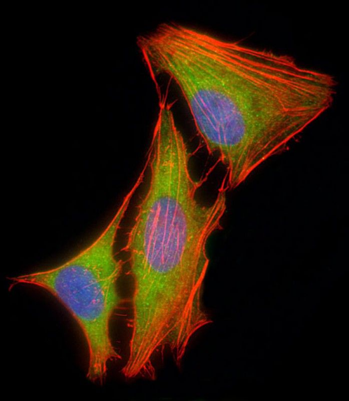
Immunofluorescent analysis of 4% paraformaldehyde-fixed, 0.1% Triton X-100 permeabilized HeLa (human cervical epithelial adenocarcinoma cell line) cells labeling MAPK3/1 with 102-20274 at 1/25 dilution, followed by Dylight® 488-conjugated goat anti-mouse IgG (NH174309) secondary antibody at 1/200 dilution (green). Immunofluorescence image showing cytoplasm and nucleus staining on HeLa cell line. Cytoplasmic actin is detected with Dylight® 554 Phalloidin (OI17558410) at 1/100 dilution (red). The nuclear counter stain is DAPI (blue).
IF

Storage/Stability

2-8°C (short-term); -20°C (long-term)
Expiration:
12 months from the date of shipment when stored properly.