RayBiotech

3607 Parkway Ln Ste 200 Norcross, Georgia 30092

www.RayBiotech.com | info@raybiotech.com

770-729-2992 | +1-888-494-8555

ISO 13485:2016 cGMP

Anti-P53 (176-200aa) Antibody

Catalog #: 130-11078

Rabbit Anti-Human P53 (176-200aa) Antibody

Product Description

Specifications

Size20 µg, 100 µg, 200 µg, 500 µg, 1 mg
Estimated Lead Time1-2 business days
SpeciesHuman
Host SpeciesRabbit
Accession Number
P04637
Gene Id
7157
Gene Symbols
TP53, P53
Protein Name / Synonyms
Cellular tumor antigen p53 (Antigen NY-CO-13) (Phosphoprotein p53) (Tumor suppressor p53)
ClonalityPolyclonal
PreparationThis antibody was produced from antiserum of rabbits immunized with a synthetic immunogen P53 (176-200aa): CPHHERCSDSDGLAPPQHLIRVEGN. This antibody was produced from a rabbit immunized with this peptide conjugated with KLH. The IgG fraction was purified by Protein A affinity chromatography.
Immunogen
Synthetic immunogen P53 (176-200aa): CPHHERCSDSDGLAPPQHLIRVEGN
Recommended ApplicationsELISA (recommended work dilution= 1:5, 000)
Reconstitution
Product is supplied as a powder obtained from lyophilization of purified antibody in glycine solution. Reconstitute the antibody with sterile water to a final concentration of 1 mg/ml.
Research AreaApoptosis, Cancer, p53 Signaling, Autophagy, DNA Damage
Shipping TypeBlue ice
Storage-20°C

Description

Description

P53 (also known as protein 53 or tumor protein 53), is a tumor suppressor protein that in humans is encoded by the TP53 gene. p53 is crucial in multicellular organisms, where it regulates the cell cycle and, thus, functions as a tumor suppressor that is involved in preventing cancer. As such, p53 has been described as "the guardian of the genome" because of its role in conserving stability by preventing genome mutation. The name p53 is in reference to its apparent molecular mass: It runs as a 53-kilodalton (kDa) protein on SDS-PAGE. But, based on calculations from its amino acid residues, p53's mass is actually only 43.7 kDa. This difference is due to the high number of proline residues in the protein, which slows its migration on SDS-PAGE, thus making it appear heavier than it actually is. This effect is observed with p53 from a variety of species, including humans, rodents, frogs, and fish.

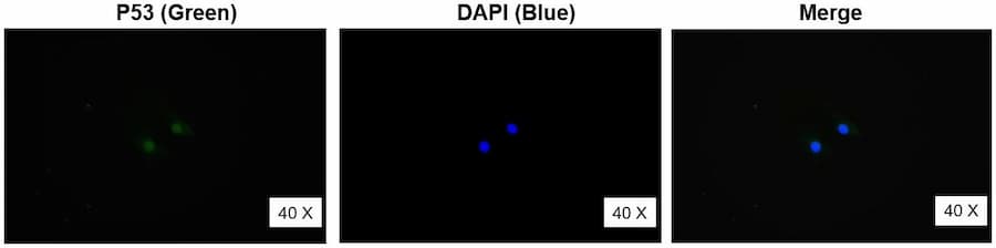
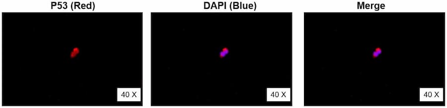
Indirect Fluorescent Antibody (IFA) Analysis

1130-11078 (1:100): Rabbit Anti-P53 (2-29aa)
Alexa Fluor 488 goat anti-rabbit (1:200)
Hela cells
Subcellular Location: nucleus, cytoplasm


1130-11078 (1:100): Rabbit Anti-P53 (2-29aa)
CY3 goat anti-rabbit (1:200)
293T cells
Subcellular Location: nucleus, cytoplasm

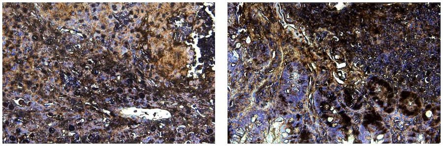
Immunohistochemistry (IHC) Analysis

IHC shows positive staining in colon cancer tumor tissue. Antibody Rabbit Anti-Human P53(2-29aa) was used at a dilution of 1/200 followed by mouse and rabbit IgG (ready to use) polymerization using HRP polymer, counterstained with hematoxylin. Heat-mediated antigen retrieval was performed using Tris/EDTA buffer pH9.0 before starting IHC staining (40X).

Storage/Stability

Store at -20°C. If intended for use within a month, may keep at 4°C. Minimize freeze-thaw cycles.

Expiration

12 months from the date of shipment when stored properly.