RayBiotech

3607 Parkway Ln Ste 200 Norcross, Georgia 30092

www.RayBiotech.com | info@raybiotech.com

770-729-2992 | +1-888-494-8555

ISO 13485:2016 cGMP

Anti-PDF Antibody

Catalog #: 144-10458

PDF Polyclonal Antibody

Product Description

Specifications

Size20 µL, 100 µL
Estimated Lead Time1-2 weeks
SpeciesHuman, Mouse, Rat
Host SpeciesRabbit
Accession Number
Q9HBH1
Gene Id
64146
Gene Symbols
PDF
Protein Name / Synonyms
PDF, peptide deformylase, mitochondrial
ClonalityPolyclonal
IsotypeIgG
Purity
Affinity purification
BuffersPBS with 0.02% sodium azide, 50% glycerol, pH7.3
Recommended ApplicationsWestern Blotting
Shipping TypeBlue ice
Storage-20°C

Description

Background

Protein synthesis proceeds after formylation of methionine by methionyl-tRNA formyl transferase (FMT) and transfer of the charged initiator f-met tRNA to the ribosome. In eubacteria and eukaryotic organelles the product of this gene, peptide deformylase (PDF), removes the formyl group from the initiating methionine of nascent peptides. In eubacteria, deformylation of nascent peptides is required for subsequent cleavage of initiating methionines by methionine aminopeptidase. The discovery that a natural inhibitor of PDF, actinonin, acts as an antimicrobial agent in some bacteria has spurred intensive research into the design of bacterial-specific PDF inhibitors. In human cells, only mitochondrial proteins have N-formylation of initiating methionines. Protein inhibitors of PDF or siRNAs of PDF block the growth of cancer cell lines but have no effect on normal cell growth. In humans, PDF function may therefore be restricted to rapidly growing cells.

Dilutions

WB 1:500 - 1:2000

Format

Calculated MW
27kDa
Observed MW
20kDa

Immunogen

Recombinant fusion protein containing a sequence corresponding to amino acids 40-243 of human PDF (NP_071736.1).

Modification

Unmodified

Storage

-20°C

Images

Western blot analysis of extracts of various cell lines, using PDF antibody (144-10458) at 1:1000 dilution.
Secondary antibody: HRP Goat Anti-Rabbit IgG (H+L) at 1:10000 dilution.
Lysates/proteins: 25ug per lane.
Blocking buffer: 3% nonfat dry milk in TBST.
Detection: ECL Basic Kit.
Exposure time: 30s.
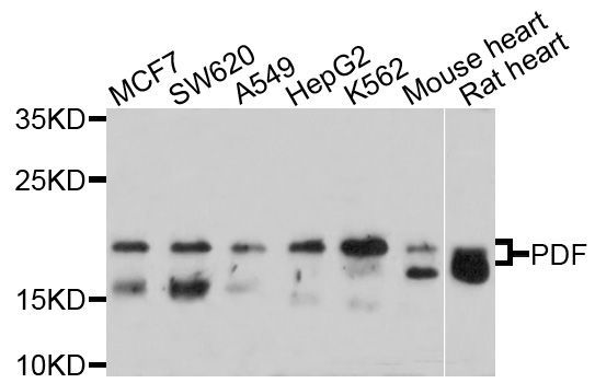
Western blot - PDF Polyclonal Antibody (144-10458)

Expiration:
12 months from the date of shipment when stored properly.