RayBiotech

3607 Parkway Ln Ste 200 Norcross, Georgia 30092

www.RayBiotech.com | info@raybiotech.com

770-729-2992 | +1-888-494-8555

ISO 13485:2016 cGMP

Anti-PDHX Antibody

Catalog #: 144-06426

PDHX Polyclonal Antibody

Product Description

Specifications

Size20 µL, 100 µL
Estimated Lead Time1-2 weeks
SpeciesHuman, Mouse, Rat
Host SpeciesRabbit
Accession Number
O00330
Gene Id
8050
Gene Symbols
PDHX
Protein Name / Synonyms
PDHX, DLDBP, E3BP, OPDX, PDX1, proX, pyruvate dehydrogenase complex component X
ClonalityPolyclonal
IsotypeIgG
Purity
Affinity purification
BuffersPBS with 0.02% sodium azide, 50% glycerol, pH7.3
Recommended ApplicationsWestern Blotting, IF
Shipping TypeBlue ice
Storage-20°C

Description

Background

The pyruvate dehydrogenase (PDH) complex is located in the mitochondrial matrix and catalyzes the conversion of pyruvate to acetyl coenzyme A. The PDH complex thereby links glycolysis to Krebs cycle. The PDH complex contains three catalytic subunits, E1, E2, and E3, two regulatory subunits, E1 kinase and E1 phosphatase, and a non-catalytic subunit, E3 binding protein (E3BP). This gene encodes the E3 binding protein subunit; also known as component X of the pyruvate dehydrogenase complex. This protein tethers E3 dimers to the E2 core of the PDH complex. Defects in this gene are a cause of pyruvate dehydrogenase deficiency which results in neurological dysfunction and lactic acidosis in infancy and early childhood. This protein is also a minor antigen for antimitochondrial antibodies. These autoantibodies are present in nearly 95% of patients with the autoimmune liver disease primary biliary cirrhosis (PBC). In PBC, activated T lymphocytes attack and destroy epithelial cells in the bile duct where this protein is abnormally distributed and overexpressed. PBC eventually leads to cirrhosis and liver failure. Alternative splicing results in multiple transcript variants encoding distinct isoforms.

Dilutions

WB 1:500 - 1:2000
IF 1:10 - 1:100

Format

Calculated MW
29kDa/51kDa/54kDa
Observed MW
54kDa

Immunogen

Recombinant fusion protein containing a sequence corresponding to amino acids 1-300 of human PDHX (NP_003468.2).

Modification

Unmodified

Storage

-20°C

Images

Western blot analysis of extracts of various cell lines, using PDHX antibody (144-06426) at 1:1000 dilution.
Secondary antibody: HRP Goat Anti-Rabbit IgG (H+L) at 1:10000 dilution.
Lysates/proteins: 25ug per lane.
Blocking buffer: 3% nonfat dry milk in TBST.
Detection: ECL Basic Kit.
Exposure time: 5s.
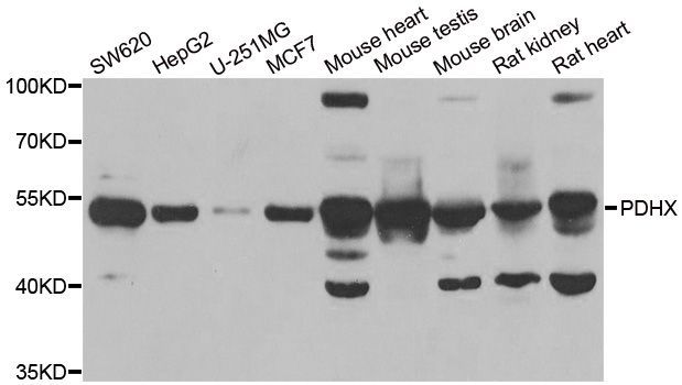
Western blot - PDHX Polyclonal Antibody (144-06426)

Immunofluorescence analysis of MCF7 cells using PDHX antibody (144-06426). Blue: DAPI for nuclear staining.
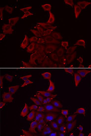
Immunofluorescence - PDHX Polyclonal Antibody (144-06426)

Expiration:
12 months from the date of shipment when stored properly.