Rabbit Anti-Human PDIA3 (C-term) Antibody, 400 ul
Loading...
Loading...
Loading...
Loading...
| Size | 400 µL |
| Estimated Lead Time | 1-2 weeks |
| Species | Human |
| Host Species | Rabbit |
| Accession Number | P30101 |
| Gene Id | 2923 |
| Gene Symbols | PDIA3 |
| Protein Name / Synonyms | PDIA3 (C-term) |
| Clonality | Polyclonal |
| Preparation | This PDIA3 antibody is generated from rabbits immunized with a KLH conjugated synthetic peptide between 446-475 amino acids from the C-terminal region of human PDIA3. |
| Concentration (lot specific) | 2.0 mg/ml |
| Recommended Applications | Western Blotting, IHC-P, FC |
| Shipping Type | Blue ice |
| Storage | -20°C |
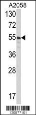 | Western blot analysis of PDIA3 Antibody (C-term) (Cat# 102-15229) in A2058 cell line lysates (35ug/lane). PDIA3 (arrow) was detected using the purified Pab. |
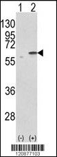 | Western blot analysis of PDIA3 (arrow) using rabbit polyclonal PDIA3 Antibody (C-term) (Cat# 102-15229). 293 cell lysates (2 ug/lane) either nontransfected (Lane 1) or transiently transfected with the PDIA3 gene (Lane 2) . |
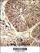 | Formalin-fixed and paraffin-embedded human lung carcinoma reacted with PDIA3 Antibody (C-term), which was peroxidase-conjugated to the secondary antibody, followed by DAB staining. This data demonstrates the use of this antibody for immunohistochemistry; clinical relevance has not been evaluated. |
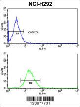 | PDIA3 Antibody (C-term) (Cat# 102-15229) flow cytometric analysis of NCI-H292 cells (bottom histogram) compared to a negative control cell (top histogram).FITC-conjugated goat-anti-rabbit secondary antibodies were used for the analysis. |
Your cart is empty.
Anti-PDIA3 (C-term) Antibody
Anti-PDIA3 (C-term) Antibody