3607 Parkway Ln Ste 200 Norcross, Georgia 30092
www.RayBiotech.com | info@raybiotech.com
770-729-2992 | +1-888-494-8555
ISO 13485:2016 cGMP

3607 Parkway Ln Ste 200 Norcross, Georgia 30092
www.RayBiotech.com | info@raybiotech.com
770-729-2992 | +1-888-494-8555
ISO 13485:2016 cGMP
Catalog #: 102-16501
Rabbit Anti-PGD (Center) Antibody, 400 ul
| Size | 400 µL |
| Estimated Lead Time | 1-2 weeks |
| Species | Human, Mouse |
| Host Species | Rabbit |
| Accession Number | P52209 |
| Gene Id | 5226 |
| Gene Symbols | PGD |
| Protein Name / Synonyms | PGD (Center) |
| Clonality | Polyclonal |
| Preparation | This PGD antibody is generated from rabbits immunized with a KLH conjugated synthetic peptide between 236-265 amino acids from the Central region of human PGD. |
| Concentration (lot specific) | 0.5 mg/ml |
| Recommended Applications | Western Blotting, IHC-P, FC |
| Shipping Type | Blue ice |
| Storage | -20°C |
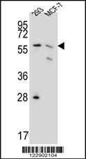 | PGD Antibody (Center) (Cat# 102-16501) western blot analysis in 293,MCF-7 cell line lysates (35ug/lane).This demonstrates the PGD antibody detected PGD protein (arrow). |
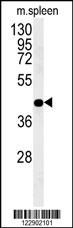 | PGD Antibody (Center) (Cat# 102-16501) western blot analysis in mouse spleen tissue lysates (35ug/lane).This demonstrates the PGD antibody detected PGD protein (arrow). |
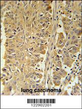 | PGD Antibody (Center) (Cat# 102-16501) immunohistochemistry analysis in formalin fixed and paraffin embedded human lung carcinoma followed by peroxidase conjugation of the secondary antibody and DAB staining. This data demonstrates the use of the PGD Antibody (Center) for immunohistochemistry. Clinical relevance has not been evaluated. |
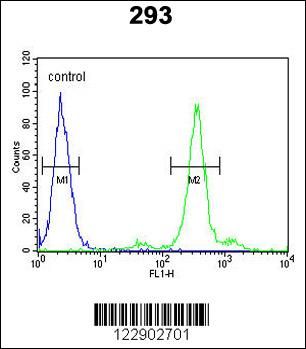 | PGD Antibody (Center) (Cat# 102-16501) flow cytometric analysis of 293 cells (right histogram) compared to a negative control cell (left histogram).FITC-conjugated goat-anti-rabbit secondary antibodies were used for the analysis. |
Your cart is empty.