3607 Parkway Ln Ste 200 Norcross, Georgia 30092
www.RayBiotech.com | info@raybiotech.com
770-729-2992 | +1-888-494-8555
ISO 13485:2016 cGMP

3607 Parkway Ln Ste 200 Norcross, Georgia 30092
www.RayBiotech.com | info@raybiotech.com
770-729-2992 | +1-888-494-8555
ISO 13485:2016 cGMP
Catalog #: 144-50304
Phospho-AKT1-T308 Polyclonal Antibody
| Size | 20 µL, 100 µL |
| Estimated Lead Time | 1-2 weeks |
| Species | Human, Mouse, Rat |
| Host Species | Rabbit |
| Accession Number | P31749 |
| Gene Id | 207 |
| Gene Symbols | AKT1 |
| Protein Name / Synonyms | AKT1, AKT, CWS6, PKB, PKB-ALPHA, PRKBA, RAC, RAC-ALPHA, AKT serine/threonine kinase 1 |
| Isotype | IgG |
| Purity | Affinity purification |
| Buffers | PBS with 0.02% sodium azide, 50% glycerol, pH7.3 |
| Recommended Applications | Western Blotting, IF |
| Research Area | Angiogenesis, Apoptosis, Inflammation, Autophagy |
| Shipping Type | Blue ice |
| Storage | -20°C |
Western blot analysis of extracts of HeLa cells, using Phospho-AKT1-T308 antibody (144-50304) at 1:2000 dilution. HeLa cells were treated by 10% FBS for after serum-starvation overnight.
Secondary antibody: HRP Goat Anti-Rabbit IgG (H+L) at 1:10000 dilution.
Lysates/proteins: 25ug per lane.
Blocking buffer: 3% BSA.
Detection: ECL Enhanced Kit.
Exposure time: 90s.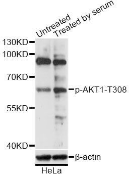
Immunofluorescence analysis of HeLa cells using Phospho-AKT1-T308 antibody (144-50304) at dilution of 1:100. Blue: DAPI for nuclear staining.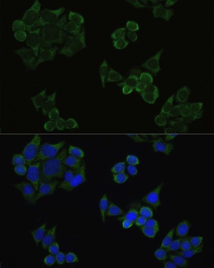
Your cart is empty.