Rabbit Anti-Phospho-IRAK1(S376) Antibody, 200 µl
Loading...
Loading...
Loading...
Loading...
| Size | 200 µL |
| Estimated Lead Time | 1-2 weeks |
| Species | Human |
| Host Species | Rabbit |
| Accession Number | P51617 |
| Gene Id | 3654 |
| Gene Symbols | IRAK1 |
| Protein Name / Synonyms | Interleukin-1 receptor-associated kinase 1, IRAK-1, 2.7.11.1, IRAK1, IRAK |
| Clonality | Polyclonal |
| Preparation | This Phospho-IRAK1(S376) antibody is generated from a rabbit immunized with a KLH conjugated synthetic peptide between 348-381 amino acids from human IRAK1. |
| Isotype | Kappa, Rabbit |
| Concentration (lot specific) | 0.5 mg/ml |
| Buffers | Purified polyclonal antibody supplied in PBS with 0.09% (W/V) sodium azide |
| Recommended Applications | IHC-P, IF, Western Blotting |
| Shipping Type | Blue ice |
| Storage | -20°C |
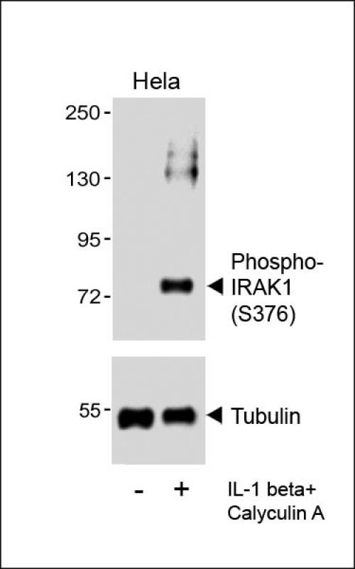 | Western blot analysis of lysates from Hela cell line, untreated or treated with IL-1 beta(20ng/ml) + Calyculin A(100nM), using (Cat. #102-18524)(upper) or Tubulin (lower). |
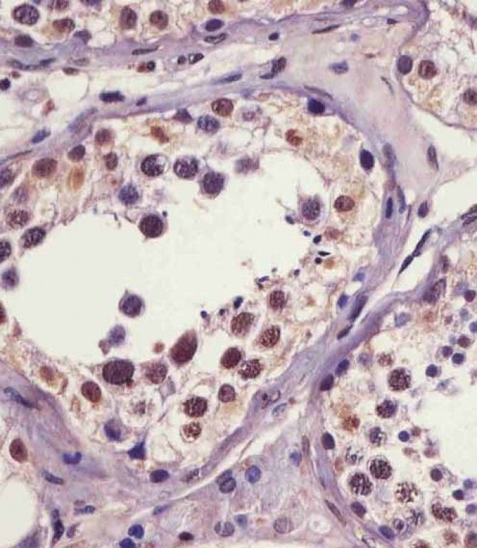
 | 102-18524 staining IRAK1 in human testis tissue sections by Immunohistochemistry (IHC-P - paraformaldehyde-fixed, paraffin-embedded sections). Tissue was fixed with formaldehyde and blocked with 3% BSA for 0. 5 hour at room temperature; antigen retrieval was by heat mediation with a citrate buffer (pH6). Samples were incubated with primary antibody (1/25) for 1 hours at 37°C. An undiluted biotinylated goat polyvalent antibody was used as the secondary antibody. |
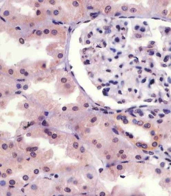
 | 102-18524 staining IRAK1 in human kidney tissue sections by Immunohistochemistry (IHC-P - paraformaldehyde-fixed, paraffin-embedded sections). Tissue was fixed with formaldehyde and blocked with 3% BSA for 0. 5 hour at room temperature; antigen retrieval was by heat mediation with a citrate buffer (pH6). Samples were incubated with primary antibody (1/25) for 1 hours at 37°C. A undiluted biotinylated goat polyvalent antibody was used as the secondary antibody. |
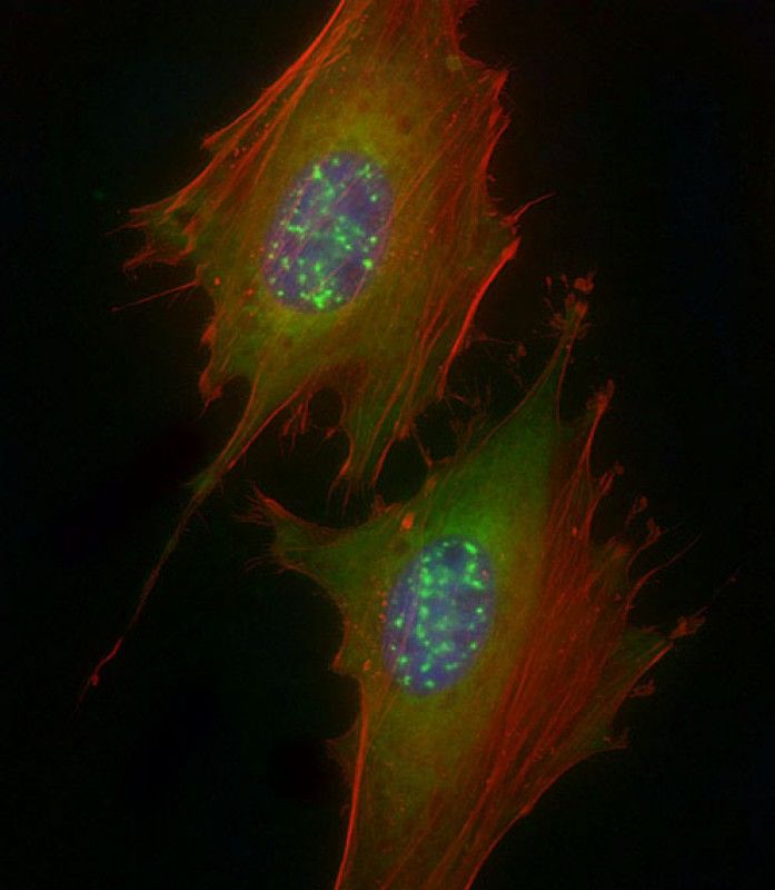 | Immunofluorescent analysis of 4% paraformaldehyde-fixed, 0.1% Triton X-100 permeabilized HeLa (human cervical epithelial adenocarcinoma cell line) cells labeling IRAK1 with 102-18525 at 1/25 dilution, followed by Dylight 488-conjugated goat anti-rabbit IgG (NK179883) secondary antibody at 1/200 dilution (green). Immunofluorescence image showing cytoplasm and nuclear speckles staining on HeLa cell line. Cytoplasmic actin is detected with Dylight 554 Phalloidin (PD18466410) at 1/100 dilution (red). The nuclear counter stain is DAPI (blue). |
Your cart is empty.
Anti-Phospho-IRAK1 (S376) Antibody
Anti-Phospho-IRAK1 (S376) Antibody