3607 Parkway Ln Ste 200 Norcross, Georgia 30092
www.RayBiotech.com | info@raybiotech.com
770-729-2992 | +1-888-494-8555
ISO 13485:2016 cGMP

3607 Parkway Ln Ste 200 Norcross, Georgia 30092
www.RayBiotech.com | info@raybiotech.com
770-729-2992 | +1-888-494-8555
ISO 13485:2016 cGMP
Catalog #: 102-15375
Rabbit Anti-Human Phospho-LC3C(S12) Antibody, 400 ul
| Size | 400 µL |
| Estimated Lead Time | 1-2 weeks |
| Species | Human |
| Host Species | Rabbit |
| Accession Number | Q9H492 |
| Gene Id | 84557 |
| Gene Symbols | MAP1LC3A |
| Protein Name / Synonyms | Phospho-LC3C(S12) |
| Clonality | Polyclonal |
| Preparation | This LC3C Antibody is generated from rabbits immunized with a KLH conjugated synthetic phosphopeptide corresponding to amino acid residues surrounding S12 of human LC3C. |
| Concentration (lot specific) | 0.5 mg/ml |
| Recommended Applications | Western Blotting, Dot Blotting |
| Research Area | Autophagy |
| Shipping Type | Blue ice |
| Storage | -20°C |
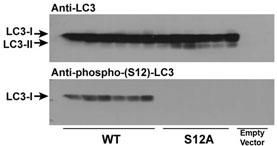 | Immunoblots of phosphorylated LC3 (phospho-LC3) in CHO cell culture. LC3 and LC3 S12A mutant vectors were transfected into CHO cells. The cell lysates were separated with SDS-PAGE and blotted with anti-phospho-LC3 S12 antibody. LC3 = microtubule-associated protein light chain-3; S12A = replacement of the amino acid position 12 serine of LC3 with alanine. WT = wildtype LC3-transfected cell lysates; S12A = LC3 S12A mutant-transfected cell lysates; Empty vector = vector with no LC3 gene. Molecular size: LC3-I = 16kDa, and LC3-II = 14 kDa |
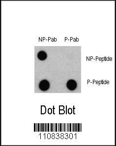 | Dot blot analysis of Phospho-LC3 (APG8a) - S12 Antibody (Cat# 102-15375) and Nonphospho-LC3 (APG8a) Antibody on nitrocellulose membrane. 50ng of Phospho-peptide or Non Phospho-peptide per dot were adsorbed. Antibody working concentrations are 0.5ug per ml. |
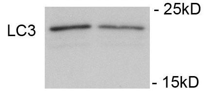 | Immunoblots of SH-SY5Y cells treated with rapamycin for 1 h was probed with 102-15375. The data shows that treatment with rapamycin showed no significant change in level of LC3. |
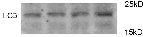 | Immunoblots of SH-SY5Y cells treated with MPP+ for 24h was probed with 102-15375. The data shows that treatment with MPP+ showed no significant change in level of LC3. |
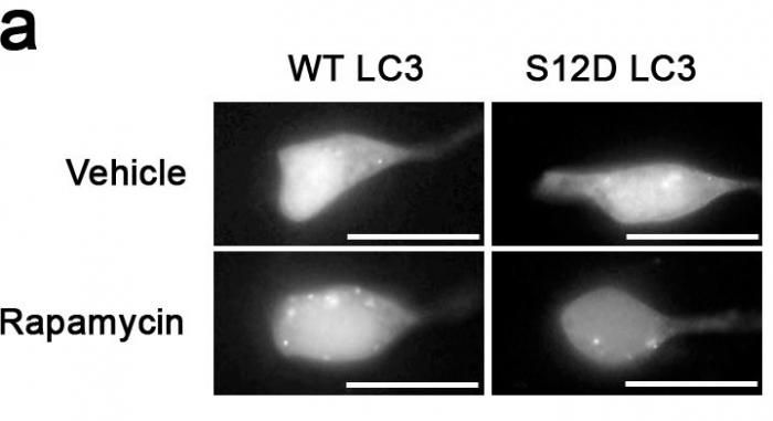 | Something like SH-SY5Y cells expressing GFP-LC3-WT or-S12D treated with rapamycin or vehicle for 1h. |
Your cart is empty.