3607 Parkway Ln Ste 200 Norcross, Georgia 30092
www.RayBiotech.com | info@raybiotech.com
770-729-2992 | +1-888-494-8555
ISO 13485:2016 cGMP

3607 Parkway Ln Ste 200 Norcross, Georgia 30092
www.RayBiotech.com | info@raybiotech.com
770-729-2992 | +1-888-494-8555
ISO 13485:2016 cGMP
Catalog #: 102-15348
Rabbit Anti-Human Phospho-Rb(S780) Antibody, 400 ul
| Size | 400 µL |
| Estimated Lead Time | 1-2 weeks |
| Species | Human |
| Host Species | Rabbit |
| Accession Number | P06400 |
| Gene Id | 5925 |
| Gene Symbols | RB1 |
| Protein Name / Synonyms | Phospho-Rb(S780) |
| Clonality | Polyclonal |
| Preparation | This Rb Antibody is generated from rabbits immunized with a KLH conjugated synthetic phosphopeptide corresponding to amino acid residues surrounding S780 of human Rb. |
| Concentration (lot specific) | 0.430 mg/ml |
| Recommended Applications | Dot Blotting, IHC-P |
| Research Area | Cancer, Inflammation |
| Shipping Type | Blue ice |
| Storage | -20°C |
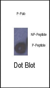 | Dot blot analysis of anti-Phospho-Rb-S780 Antibody (Cat# 102-15348) on nitrocellulose membrane. 50ng of Phospho-peptide or Non Phospho-peptide per dot were adsorbed. Antibody working concentrations are 0.5ug per ml. |
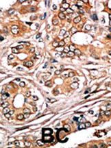 | Formalin-fixed and paraffin-embedded human cancer tissue reacted with the primary antibody, which was peroxidase-conjugated to the secondary antibody, followed by AEC staining. This data demonstrates the use of this antibody for immunohistochemistry; clinical relevance has not been evaluated. BC = breast carcinoma; HC = hepatocarcinoma. |
Your cart is empty.