Rabbit Anti-Human PI3KCD (C-term) Antibody, 400 ul
Loading...
Loading...
Loading...
Loading...
| Size | 400 µL |
| Estimated Lead Time | 1-2 weeks |
| Species | Human |
| Host Species | Rabbit |
| Accession Number | O00329 |
| Gene Id | 5293 |
| Gene Symbols | PIK3CD |
| Protein Name / Synonyms | PI3KCD (C-term) |
| Clonality | Polyclonal |
| Preparation | This PI3KCD antibody is generated from rabbits immunized with a KLH conjugated synthetic peptide between 687-717 amino acids from the C-terminal region of human PI3KCD. |
| Concentration (lot specific) | 2.0 mg/ml |
| Recommended Applications | Western Blotting, IHC-P, IF |
| Research Area | Angiogenesis, Inflammation |
| Shipping Type | Blue ice |
| Storage | -20°C |
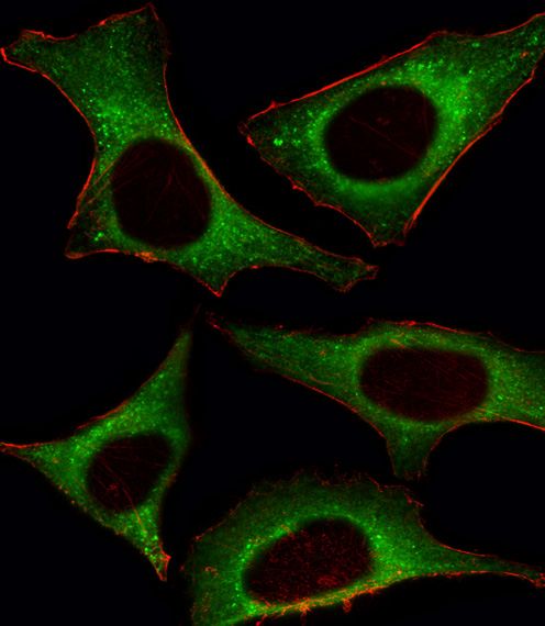 | Fluorescent image of Hela cell stained with PI3KCD Antibody (C-term)(Cat# 102-17760).Hela cells were fixed with 4% PFA (20 min), permeabilized with Triton X-100 (0.1%, 10 min), then incubated with PI3KCD primary antibody (1:25, 1 h at 37°). For secondary antibody, Alexa Fluor® 488 conjugated donkey anti-rabbit antibody (green) was used (1:400, 50 min at 37°).Cytoplasmic actin was counterstained with Alexa Fluor® 555 (red) conjugated Phalloidin (7units/ml, 1 h at 37°).PI3KCD immunoreactivity is localized to Cytoplasm significantly. |
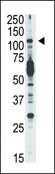 | Western blot analysis of anti-PI3KCD Pab (Cat# 102-17760) in Jurkat cell lysate. PI3KCD (arrow) was detected using purified Pab. Secondary HRP-anti-rabbit was used for signal visualization with chemiluminescence. |
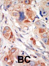 | Formalin-fixed and paraffin-embedded human cancer tissue reacted with the primary antibody, which was peroxidase-conjugated to the secondary antibody, followed by DAB staining. This data demonstrates the use of this antibody for immunohistochemistry; clinical relevance has not been evaluated. BC = breast carcinoma; HC = hepatocarcinoma. |
Your cart is empty.
Anti-PI3KCD (C-term) Antibody
Anti-PI3KCD (C-term) Antibody