RayBiotech

3607 Parkway Ln Ste 200 Norcross, Georgia 30092

www.RayBiotech.com | info@raybiotech.com

770-729-2992 | +1-888-494-8555

ISO 13485:2016 cGMP

Anti-SIGLEC7 (D-siglec) (N-term) Antibody

Catalog #: 102-22234

Rabbit Anti-SIGLEC7 (D-siglec) (N-term) Antibody, 400 µl

Product Description

Specifications

Size400 µL
Estimated Lead Time1-2 weeks
SpeciesHuman
Host SpeciesRabbit
Accession Number
Q9Y286
Gene Id
27036
Gene Symbols
SIGLEC7
Protein Name / Synonyms
Sialic acid-binding Ig-like lectin 7, Siglec-7, Adhesion inhibitory receptor molecule 1, AIRM-1, CDw328, D-siglec, QA79 membrane protein, p75, CD328, SIGLEC7, AIRM1
ClonalityPolyclonal
CloneRB2462
IsotypeKappa, Rabbit
Recommended ApplicationsWestern Blotting, IHC-P
Shipping TypeBlue ice
Storage-20°C

Description

Introduction

SIGLECs are members of the immunoglobulin superfamily that are expressed on the cell surface. Most SIGLECs have one or more cytoplasmic immune receptor tyrosine-based inhibitory motifs (ITIM). SIGLECs are typically expressed on cells of the innate immune system, with the exception of the B-cell expressed SIGLEC6. Sequence analysis predicted that the 697-amino acid SIGLEC10 protein contains a signal peptide, an N-terminal V-set Ig-like domain and four C2-set Ig-like domains, five potential N-linked glycosylation sites, a transmembrane region, and a 126-residue cytoplasmic tail with 3 putative ITIMs. Northern blot analysis detected a major 3.0-kb SIGLEC10 transcript, with highest levels in spleen, lymph node, blood leukocytes, and appendix. Little or no expression was observed in pancreas, thyroid, and testis. Flow cytometric analysis demonstrated eosinophil-specific expression of SIGLEC10, but at a lower level than that of SIGLEC8. Expression was also detected on monocytes and a CD16-positive/CD56-negative natural killer-like lymphocyte population. After sialidase treatment, which is necessary for unmasking the sialic acid-binding site on SIGLECs interacting with cell surface sialic acids, cells expressing SIGLEC10 bound to red blood cells. Immunoprecipitation analysis indicated expression of a 100- to 120-kD monomeric protein, higher than the predicted molecular mass, suggesting that SIGLEC10 is glycosylated.

Other Information

NCBI Accession #
NP_001264130.1;NP_055200.1;NP_057627.2
Antigen Type
Synthetic Peptide
Format
Purified polyclonal antibody supplied in PBS with 0.09% (W/V) sodium azide. This antibody is prepared by Saturated Ammonium Sulfate (SAS) precipitation followed by dialysis against PBS.
Calculated Molecular Weight (Da)
51143
Antigen Source
Human

Images

WB (1:1000)

All lanes : Anti-SIGLEC7 Antibody (W20) at 1:1000 dilution Lane 1: human liver lysate Lane 2: human placenta lysate Lysates/proteins at 20 µg per lane. Secondary Goat Anti-Rabbit IgG, (H+L), Peroxidase conjugated at 1/10000 dilution. Predicted band size : 51 kDa Blocking/Dilution buffer: 5% NFDM/TBST.
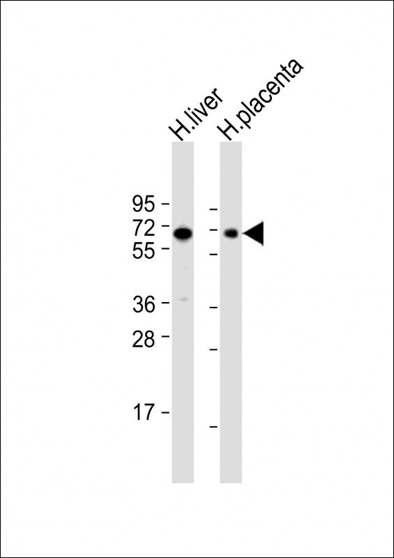
WB

Storage/Stability

2-8°C (short-term); -20°C (long-term)
Expiration:
12 months from the date of shipment when stored properly.