In December 2019, the first cases of patients with severe acute respiratory syndrome (SARS) caused by the novel coronavirus, SARS-CoV-2, were reported in China. [1] In mid-March 2020, the World Health Organization (WHO) declared COVID-19 (COronaVIrus Disease of 2019) a pandemic. By mid-May 2020, there were ~4.5 million confirmed cases and ~300,000 deaths in 227 countries worldwide. [2] In this blog, the basic biology of how the SARS-CoV-2 virus causes COVID-19 is reviewed. The symptoms, diagnosis, and current research of COVID-19 are also reviewed.
The Virus
SARS-CoV-2 is the virus responsible for COVID-19 infection. It is thought that SARS-CoV-2 was originally transmitted from bats to humans through an intermediate host because of its 98-99% similarity to two bat-derived coronaviruses, bat-SL-CoVZC45 and bat-SL-CoVZXC21. [3, 4] Surprisingly, SARS-CoV-2 has lower homology (~75%) to the SARS virus (SARS-CoV) from the 2003 outbreak. [3]
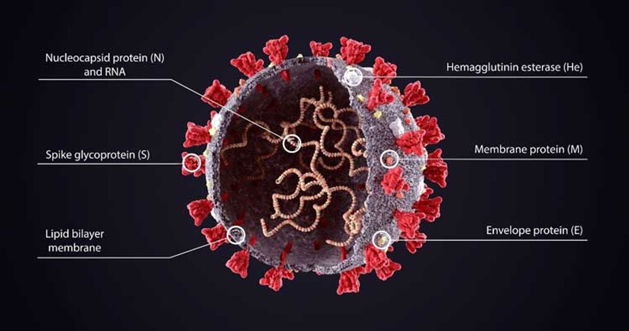
The outside of the viral particle is comprised of four main proteins: spike (S), envelope (E), membrane (M), and hemagglutinin esterase (HE) (Figure 1). [5] Inside the viral particle, the viral RNA is encased in a shell made of the nucleocapsid (N) protein. Notably, there are also 16 nonstructural proteins and 5- 8 accessory proteins. [6] The S protein is of particular interest to the research community because it is necessary for the virus to enter human cells. Other studies have demonstrated that the N and S proteins have high immunogenicity, which means that they are often targeted by antibodies produced by the patient’s immune system. [7 - 10]
Hello.
Great information thank you . I have a question , how long can you test positive for antibodies specifically the N protein.and is there any know way to help flush it out of ones system
I really appreciated your page with a very clear and nice material on COVID-19 Biology 101. Congratulations on the job. If you have more nice pictures and ilustration about COVID-19 Biology 101
I would like to receive it...GOOD JOB
PAULO A MELO MD PhD
Leave a Reply
Figure 1: Structure of the SARS-CoV-2 virus
Viral Entry
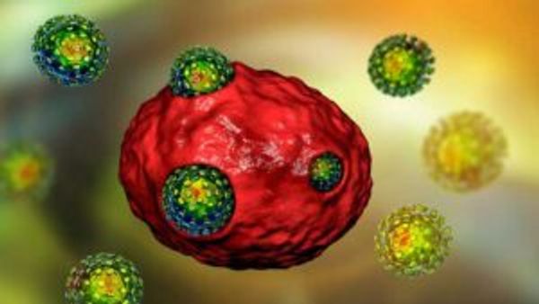
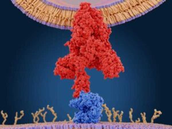
Two transmembrane proteins, ACE2 and TMPRSS2, displayed on the surface of human cells are required for SARS-CoV-2 viral entry (Figure 2). [11] First, the angiogenin I converting enzyme 2 (ACE2) receptor binds to the SARS-CoV-2 S protein via the S protein’s receptor binding domain (RBD) (Figure 3). The S protein is comprised of a signal peptide, S1 subunit containing the RBD, the S2 subunit, and a transmembrane domain (Figure 4). [12] Interestingly, the S1 subunit shares little homology with the SARS-CoV spike protein whereas the S2 subunit is nearly identical (89.8%). [13] A recent study revealed that the SARS-CoV-2 RBD binds to ACE2 with higher binding affinity than the SARS-CoV RBD, which may explain why SARS-CoV-2 is more contagious than SARS-CoV. [13, 14] The ACE2 receptor's known biological functions include vasodilation and amino acid transport. [15, 16] It is expressed in a wide variety of tissues, including the lungs, small intestine, and adipose tissue. [17]
Figure 2. Viral entry of the SARS-CoV-2 virus into a human cell (red)
Figure 3. Interaction between the SARS-CoV-2 spike protein (red) and human ACE2 receptor (blue)
Second, the transmembrane serine protease 2 (TMPRSS2) primes the S protein for import into the endoplasmic reticulum (ER) by cleaving the S protein twice: once between the S1 and S2 subunits and once within the S2 subunit. [18] Within the ER, the S protein is highly glycosylated, which may contribute to immune evasion. [19 - 21] TMPRSS2's specific biological functions are unknown in human cells, but the protein is upregulated in cancer and contributes to cancer cell invasion and metastasis. [22]
COVID-19 Symptoms
A wide range of symptoms of COVID-19 appearing 2 – 14 days after exposure have been reported. The symptoms include fever, cough, headache, muscle pain, chills, sore throat, pernio-like lesions of the toes, and loss of taste or smell. While many patients are asymptomatic or have mild symptoms, 4.6% of diagnosed patients are hospitalized with 7.1% of admitted patients ending in death. [23] It is important to note that COVID-19 patients displaying little to no symptoms are still contagious. Moreover, advanced age and underlying conditions are associated with higher morbidity and mortality rates. In a recent study by the Centers for Disease Control (CDC), 7.4% and 13.8% of diagnosed adults in the United States 50 – 64 years and > 65 years are hospitalized, respectively. Of the COVID-19 related deaths in the United States, 0.9%, 7.0%, 12.5%, and 79.6% have been attributed to patients that are < 34, 35 – 54, 55 – 64, and > 65 years old, respectively. [24]
SARS-CoV-2 mainly attacks cells in the respiratory system, possibly because SARS-CoV-2 is usually spread through respiratory droplets or touching the mouth with contaminated hands. [25] This can lead to acute respiratory distress syndrome in severe cases. However, systemic organ and tissue damage other than the lungs (e.g., heart, kidneys, liver) can also occur. How the virus causes tissue and organ damage – whether directly or indirectly – remains unclear.
Immune Response
COVID-19 infection does not go unnoticed by the immune system. One likely explanation for the tissue and organ damage that occurs during COVID-19 is a "cytokine storm," which is an out-of-control immune response to SARS-CoV-2 during which a multitude of inflammatory proteins are released at once. [26] While SARS-CoV-2 stimulates the immune response, it also blocks the release of interferons that are important in cellular defenses against viral propagation, which is normally achieved by stopping or decreasing cellular metabolism, protein transport, and transcription. [27] Importantly, host cell transcription is hijacked by the virus to replicate more of itself. Indeed, high levels of inflammatory markers have been associated with COVID-19 severity and prognosis.
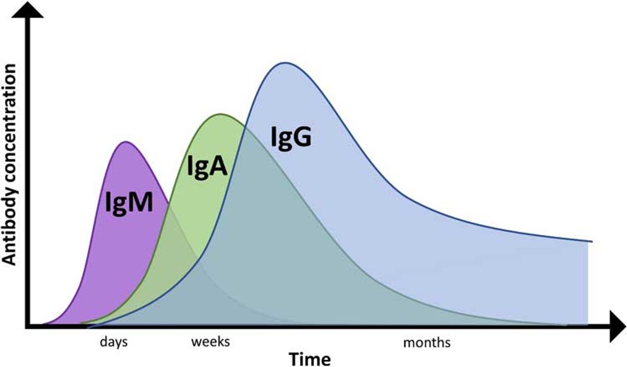
Antibodies to SARS-CoV-2 proteins are also produced. The first antibody isotype that is generated is IgM, which reflects acute infection. [28] Within days to weeks, the short-lived, low-affinity IgM antibodies are converted to long-lived, high-affinity IgG and IgA antibodies (Figure 5). IgG antibodies are especially high in blood, whereas IgA antibodies are high at mucosal surfaces and in secretions. A study of 208 COVID-19 patients revealed that IgM and IgA antibodies could be first detected within 3 – 6 days whereas IgG antibodies could be detected 10 – 18 days after symptom onset. [29]
Figure 5. General antibody profile across time following infection. Antibody profiles vary across individuals and health status.
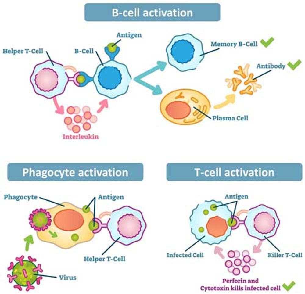
Helper T cells are important in the adaptive immune response because they help activate B cells, phagocytes, and killer T cells to target or kill infected cells (Figure 6). [30] Two studies using flow cytometry showed that 25 of the 28 patients who had recovered from COVID-19 carried helper T cells that recognized the S protein. [31, 32] Other SARS-CoV-2 proteins were also recognized by their helper T cells. Interestingly, 23 of 68 (34%) uninfected people also produced helper T cells that recognized SARS-CoV-2 likely due to previous coronavirus infections, and could confer some protection from COVID-19 infection.
Figure 6. Helper T cells play essential roles in the adaptive immune response
COVID-19 Diagnosis
COVID-19 status is determined based on clinical symptoms (described above), viral load, and the presence of antibodies to SARS-CoV-2 proteins. A patient displaying COVID-19 symptoms is not enough for diagnosis as the physical manifestations can occur from other types of infections. Thus, a physician will rely on two platforms to confirm COVID-19:
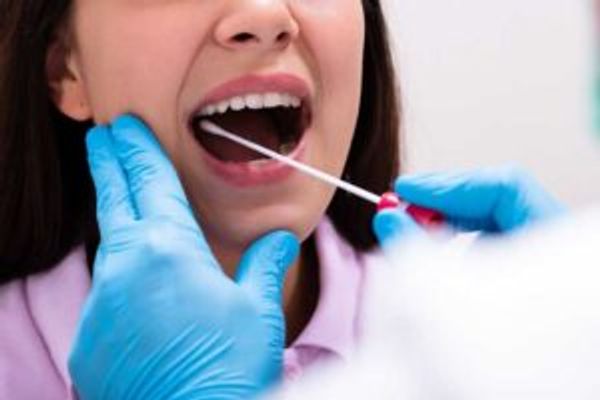
Reverse transcription polymerase chain reaction (RT-PCR): SARS-CoV-2 virus is detected with this technique. To do this, the back of the throat or nasal cavity is swabbed and sent to a laboratory for analysis (Figure 7). The viral RNA from the swab is extracted and subjected to RT-PCR, which amplifies the viral RNA for detection. Quantitative results are obtained within 2 – 4 days. This technique is considered the gold standard test; however, high false negative rates (> 20%) have been reported. [33, 34] The high false negative rate may be due to improper sample collection (e.g., not going far enough back in the nasal cavity). These tests must be performed in a high complexity clinical laboratory improvement amendments (CLIA) laboratory. Of note, patients who have recovered from COVID-19 and no longer have viral RNA cannot use this test to confirm that they had COVID-19.
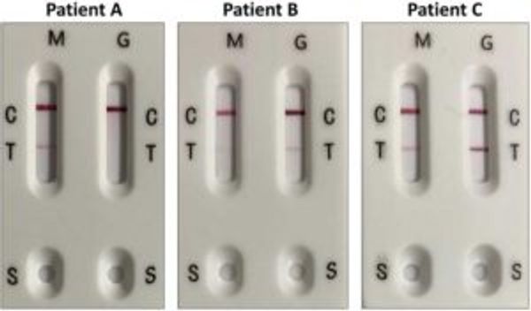
Antibody tests: Different antibody isotypes (IgM, IgG) to SARS-CoV-2 proteins in the blood can be detected in different ways. The first relies on enzyme-linked immunosorbent assay (ELISA) technology. Here, a SARS-CoV-2 protein is immobilized in a well of multi-well plate. Serum or plasma is added to the well, during which antibodies that target the SARS-CoV-2 protein will bind. Unbound sample will be washed off and the captured antibody is bound and detected using a labeled anti-human antibody that changes the color of the well when a specific substrate is added. These types of tests are performed by CLIA laboratories and require a compatible plate reader. The turnaround time is 1 – 4 days.Rapid serology tests detect COVID-19 antibodies in less than 10 minutes using lateral flow technology similar to a home pregnancy test (Figure 8). Briefly, a drop of serum, whole blood, or finger prick samples are mixed with sample diluent, and then 2 – 3 drops of the diluted sample is placed into the inlet of the hand-held test. The antibodies will migrate across a strip on which a SARS-CoV-2 protein is immobilized. If antibodies to that protein are present, they will bind to the protein and the antibodies are detected via a color change visual to the naked eye (Figure 8). See a video on how to use one here. Advantages of these tests are that the turnaround time is short and no instrumentation is required. There are different types rapid tests available, from how the test is constructed to how the test should be performed. Notably, no rapid test has been approved for at-home use. Rather, these tests must be performed either by a medical professional or under the umbrella of a high-complexity CLIA laboratory.Like the PCR-based test, high false negative rates have also been reported with antibody tests. [34] False negatives can occur for numerous reasons, but the primary cause is that the antibody level is below the detection limit of the test. Antibodies may not be detected because the sample was collected too early or too late based on the patient’s unique antibody profile across time. For this reason, both IgM and IgG antibodies should be tested to decrease the false negative rate (Figures 5, 8). Another reason why antibodies may not be detected is that the patient did not generate antibodies to the specific SARS-CoV-2 protein(s) that the test is detecting. Rather, a patient may have developed antibodies to other SARS-CoV-2 proteins. Since COVID-19 primarily affects the respiratory system, the analysis of IgA antibodies in saliva or the nasal cavity has increased.
Figure 7. The back of the throat or nasal cavity is swabbed for SARS-CoV-2 viral RNA detection
Figure 8. IgM and IgG antibody detection in three COVID-19 patients with a rapid antibody serology test. The rapid test here detects antibodies to the SARS-CoV-2 N protein. Patient A has IgM antibodies, but not IgG antibodies to the N protein. Patients B and C have both IgM and IgG antibodies to the N protein, but at varying levels. M = IgM. G = IgM. C = Control line, a positive control band that should always be present. T = Test line, presence of a pink-hued band regardless of saturation level indicates COVID-19 infection. S = Sample bed where sample is first placed.
To improve the accurate diagnosis of COVID-19 patients, both PCR and antibody-based tests should be performed.
Roles of Neutralizing Molecules in COVID-19 Treatment & Prevention
Neutralizing molecules that reduce viral infectivity can be used to treat and prevent COVID-19. These molecules can be chemically- or biologically-derived. Thus, a major focus of COVID-19 research is identifying molecules that block the binding between the SARS-CoV-2 S protein and the human ACE2 receptor, a critical step in viral entry (see also "Viral Entry")(Figure 2). High throughput S-ACE2 binding assays have the advantage of screening potential neutralizing molecules – from small molecule inhibitors to patient serum – rapidly. Neutralizing molecules can also be studied in culture, either with live SARS-CoV-2 or with a SARS-CoV-2 pseudovirus, which is a virus that can mimic viral entry but is not considered a live virus. Some vaccines, such as the vaccine to seasonal flu [34], are designed to elicit the immune response to generate neutralizing antibodies to the virus. Understanding which SARS-CoV-2 proteins or protein regions are highly antigenic is crucial in vaccine development. In one study, the analysis of serological antibodies revealed that the most antigenic regions specific to COVID-19 patients were the S protein’s RBD and extracellular domain. [8] Other studies show that the S protein is the most immunogenic SARS-CoV-protein. [9, 10] Antibody profiling can be performed with commercially-available peptide arrays.
Conclusions
The research community has worked tenaciously and quickly to help understand the SARS-CoV-2 virus and the COVID-19 pandemic, yet questions remain. Where exactly did the virus originate? How will the virus evolve? Since testing is performed only for patients who are suspected to have COVID-19, how many people have been or are truly infected? Do recovered patients have immunity to COVID-19 and, if so, for how long? How will warm weather affect the transmission and spread of COVID-19? For the most effective vaccine, which SARS-CoV-2 proteins or protein regions should be used? What is the host proteomic response during early, acute, and recovering COVID-19 infection? To help answer some of these questions, please check out RayBiotech's comprehensive catalog of COVID-19 research products and in vitro diagnostics tests here.
For a general overview of the research tools used to detect and study COVID-19, please read our blog, "COVID-19 Research Tools 101."
References
He F, et al. Coronavirus disease 2019: What we know? J Med Virol. 2020 Mar 14.
Worldometers.info. 17 May, 2020. Dover, Delaware, U.S.A.
Lai C, et al. Severe acute respiratory syndrome coronavirus 2 (SARS-CoV-2) and coronavirus disease-2019 (COVID-19): The epidemic and the challenges. Int J Antimicrob Agents. 2020 Feb 17: 105924.
Zhou P, et al. A pneumonia outbreak associated with a new coronavirus of probable bat origin. Nature volume 579, pages270–273(2020).
Astuti I and Ysrafil. Severe Acute Respiratory Syndrome Coronavirus 2 (SARS-CoV-2): An overview of viral structure and host response. Diabetes Metab Syndr. 2020 Apr 18
Jiang S, et al. Neutralizing Antibodies against SARS-CoV-2 and Other Human Coronaviruses. Trends Immunol. 2020 May;41(5):355-359.
Ahmed SF, et al. Preliminary Identification of Potential Vaccine Targets for the COVID-19 Coronavirus (SARS-CoV-2) Based on SARS-CoV Immunological Studies. Viruses. 2020 Mar; 12(3): 254.
Zhang X, et al. Proteome-wide analysis of differentially-expressed SARS-CoV-2 antibodies in early COVID-19 infection. medRxiv preprint (published online May 2). 2020; Available from: https://www.medrxiv.org/content/10.1101/2020.04.14.20064535v2
Kontou P, et al. Antibody tests in detecting SARS-CoV-2 infection: a meta-analysis. medRxiv preprint (published online April 25). 2020; Available from: https://www.medrxiv.org/content/10.1101/2020.04.22.20074914v1.
Jiang H, et al. Global profiling of SARS-CoV-2 specific IgG/ IgM responses of convalescents using a proteome microarray. medRxiv preprint (published online March 27). 2020; Available from: https://www.medrxiv.org/content/10.1101/2020.03.20.20039495v1
Hoffmann M, et al. SARS-CoV-2 Cell Entry Depends on ACE2 and TMPRSS2 and Is Blocked by a Clinically Proven Protease Inhibitor. Cell. 2020 Apr 16;181(2):271-280.e8.
Ou X, et al. Characterization of spike glycoprotein of SARS-CoV-2 on virus entry and its immune cross-reactivity with SARS-CoV. Nat Commun. 2020; 11: 1620.
Xia S, et al. Inhibition of SARS-CoV-2 (previously 2019-nCoV) infection by a highly potent pan-coronavirus fusion inhibitor targeting its spike protein that harbors a high capacity to mediate membrane fusion. Cell Research volume 30, pages343–355(2020)
Wang Q, et al. Structural and Functional Basis of SARS-CoV-2 Entry by Using Human ACE2. 020, Cell181, 894–904
de Moraes PL, et al. Vasodilator Effect of Angiotensin-(1-7) on Vascular Coronary Bed of Rats: Role of Mas, ACE and ACE2. Protein Pept Lett. 2017 Nov 17;24(9):869-875.
Camargo SM, et al. Tissue-specific amino acid transporter partners ACE2 and collectrin differentially interact with hartnup mutations. Gastroenterology. 2009 Mar;136(3):872-82.
Li M, et al. Expression of the SARS-CoV-2 cell receptor gene ACE2 in a wide variety of human tissues. Infectious Diseases of Poverty volume 9, Article number: 45 (2020)
Hoffmann M, et al. Priming Time: How Cellular Proteases Arm Coronavirus Spike Proteins. Activation of Viruses by Host Proteases. 2018 Feb 16 : 71–98.
Chakraborti S, et al. The SARS Coronavirus S Glycoprotein Receptor Binding Domain: Fine Mapping and Functional Characterization. Virology Journal volume 2, Article number: 73 (2005)
Watanabe Y, et al. Site-specific glycan analysis of the SARS-CoV-2 spike. Science. 04 May 2020: eabb9983
Shajahan A, et al. Deducing the N- and O-glycosylation profile of the spike protein of novel coronavirus SARS-CoV-2. Glycobiology. 2020 May 4.
Lucas JM, et al. The androgen-regulated protease TMPRSS2 activates a proteolytic cascade involving components of the tumor microenvironment and promotes prostate cancer metastasis. Cancer Discov. 2014 Nov;4(11):1310-25.
Garg S, et al. Hospitalization rates and characteristics of patients hospitalized with laboratory-confirmed coronavirus disease 2019 – COVID-NET, 14 states, March 1 – 30, 2020. MMWR Morb Mortal Wkly Rep 2020; 69:458-464.
Cevik M, et al. COVID-19 pandemic - A focused review for clinicians. Clin Microbiol Infect. 2020 Apr 25.
Gen Y, et al. Pathophysiological Characteristics and Therapeutic Approaches for Pulmonary Injury and Cardiovascular Complications of Coronavirus Disease 2019. Cardiovasc Pathol. 2020 Apr 17 : 107228.
Zhao M. Cytokine storm and immunomodulatory therapy in COVID-19: role of chloroquine and anti-IL-6 monoclonal antibodies. Int J Antimicrob Agents. 2020 Apr 16 : 105982.
Blanco-Melo D, et al. Imbalanced host response to SARS-CoV-2 drives development of COVID-19. 2020, Cell 181, 1–10
Schroeder H, et al. Structure and Function of Immunoglobulins. J Allergy Clin Immunol. 2010 Feb; 125(2 0 2): S41–S52.
Guo L, et al. Profiling Early Humoral Response to Diagnose Novel Coronavirus Disease (COVID-19). Clin Infect Dis. 2020 Mar 21.
Alberts B, et al. Molecular Biology of the Cell. 4th edition. New York: Garland Science; 2002.
Grifoni A, et al. Targets of T cell responses to SARS-CoV-2 coronavirus in humans with COVID-19 disease and unexposed individuals, Cell (2020).
Braun J, et al. Presence of SARS-CoV-2 reactive T cells in COVID-19 patients and healthy donors. medRxiv preprint (published online April 22). 2020; Available from: https://www.medrxiv.org/content/10.1101/2020.04.17.20061440v1
Xiao AT, et al. False-negative of RT-PCR and prolonged nucleic acid conversion in COVID-19: Rather than recurrence. J Med Virol. 2020 Apr 9.
Dheda K, et al. Diagnosis of COVID-19: Considerations, Controversies and Challenges in South Africa. Wits Journal of Clinical Medicine. 2020 Apr; 2(SI): 3–10.
Gomez Lorenzo MM and Fenton MJ. Immunobiology of Influenza Vaccines. Chest. 2013 Feb; 143(2): 502–510.