Skip to navigationSkip to contentSkip to footer
Need Help?770-729-2992
MY ACCOUNT
CART
RayBiotech
RayBiotech
  • Products
    • Multiplex Assays
    • ELISA Kits
    • Proteins
    • Antibodies
    • Flow Cytometry
    • Assay Kits
    • Molecular Biology
    • Other Products
    • Human Cytokine Array C5
    • Human IL-6 ELISA
    • Human Inflammation Array Q3
    • Mouse Cytokine Array C3
    • Recombinant SARS-CoV-2 Spike Protein, S1 Subunit
    • Human Phosphorylation Pathway Profiling Array C55
    • Antibody Arrays
    • Proteome Profiling Arrays
    • Cytometric Bead Arrays
    • Epitope Mapping Peptide Arrays
    • Protein Arrays
    • PTM Multiplex Assays
    • NexaTag® Ultrasensitive Multiplex ELISA
    • Sandwich ELISA
    • Competitive ELISA
    • PTM ELISA
    • NexaTag® Ultrasensitive qIPCR ELISA
    • Indirect ELISA
    • Bridging ELISA
    • Cell-Based ELISA
    • Recombinant Proteins
    • GMP Proteins
    • Native Proteins
    • Peptides
    • Primary Antibodies
    • Secondary Antibodies
    • Flow Cytometry Antibodies
    • Isotype Controls
    • Antibody Pairs
    • Flow Cytometry Antibodies
    • Flow Cytometry Reagents
    • Flow Cytometry Assays
    • Cytometric Bead Arrays
    • Fluorescent Beads
    • Quantum Dots
    • Metabolism Assays
    • Ligand Binding Assays
    • Drug Antibody (DA) & ADA Assays
    • Transcription Factor Activity Assays
    • PCR Assays
    • Nucleic Acid Assays
    • Epigenetics
    • Mitochondrial Assays
    • Reagents
    • Cell Culture
    • Biospecimen Samples
    • Sample Collection & Preparation
    • Lysates
    • Magnetic Beads
    • Lab Equipment and Supplies
    • Biological Buffers

    Featured Products

    Human Cytokine Array C5Human IL-6 ELISAHuman Inflammation Array Q3Mouse Cytokine Array C3Recombinant SARS-CoV-2 Spike Protein, S1 SubunitHuman Phosphorylation Pathway Profiling Array C55
  • Services
    • Multiplex Assay Services
    • ELISA Services
    • Protein Services
    • Antibody Services
    • CRO Services
    • Flow Cytometry Services
    • SIMOA – Single Molecule Array
    • Other Services
    • Proteomic Services
    • Custom Arrays
    • Array Scanning and Analysis
    • Custom Protein Production
    • GMP Protein Production
    • IHC Controls
    • Stable Cell Line Development
    • Molecular Biology Services
    • Antibody Production
    • Recombinant Antibody Expression
    • Bulk Antibody Production
    • Antibody Conjugation Services
    • Cell Biology Services
    • Epitope Mapping Services
    • Biomarker Discovery
    • Cell Biology Services
    • Antibody Drug Development & Characterization
    • Pharmacokinetic and Pharmacodynamic Analysis
    • Assay Development Services
    • Molecular Biology Services
    • Multiomics Services
    • Diagnostic Assay Development
    • GMP Protein Production
    • Biostatistics and Bioinformatics
    • Bulk Antibody Production
    • QC Testing Services
    • Auto-Western Blot Service
    • COVID-19 Pseudovirus Service
    • Biospecimen Samples & Sample Services
    • RNA & DNA Methylation Detection Services
  • Research Areas
    • Cell Signaling Pathways
    • Areas of Interest
    • Akt Signaling Pathway
    • AMPK Signaling pathway
    • ErbB Signaling Pathway
    • Hedgehog Signaling Pathway
    • HIF1-alpha Signaling Pathway
    • IGFR1 Signaling Pathway
    • JAK-STAT Signaling Pathway
    • MAPK Signaling Pathway
    • mTOR Signaling Pathway
    • NF-kappaB Signaling Pathway
    • Notch Signaling Pathway
    • p53 Signaling Pathway
    • PKC Pathway
    • TGF-beta Signaling Pathway
    • Wnt/beta-Catenin Pathway
    • Antibody Drug Development
    • Autoimmunity & Inflammation
    • Cancer
    • Cardiovascular Disease
    • Infectious Disease & Vaccines
    • Neuroscience
    • Obesity
    • Post Translational Modifications
    • RNA & DNA Modifications
  • Resources
    • Array Picker Tool
    • Sample Shipment Instructions
    • Sample Preparation Tips
    • Publications / Citations
    • Promotions
    • Resource Library
    • Learning Center
    • Manual Protocols
    • Array Analysis Tools
  • Contact Us
    • Distributors & Service Providers
  • About Us
    • Quality Systems
    • Business Partnerships
    • Careers
    • News & Events
Exosome Services for Advanced Research
  • Home
  • Flow Cytometry Services
  • Exosome Services for Advanced Research

Exosome Services for Advanced Research

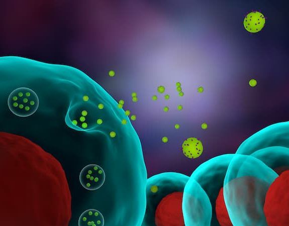
Exosomes are nanosized extracellular vesicles (30–150 nm) released by nearly all cell types and play a central role in intercellular communication. They transport functional biomolecules—including proteins, mRNA, miRNA, and lipids—to recipient cells, influencing pathways involved in tissue regeneration, immune modulation, inflammation, and disease progression. Because of their natural biocompatibility, stability, and targeting specificity, exosomes have emerged as powerful platforms for therapeutic development and drug delivery. In addition, exosomes reflect the molecular composition of their cells of origin and are readily detectable in biofluids, making them highly informative, minimally invasive biomarkers for disease diagnosis, prognosis, and treatment monitoring.

RayBiotech provides end-to-end services supporting both basic and translational research with reliable, scalable, and well-characterized solutions. Whether your work centers on mechanistic studies, diagnostic development, or therapeutic engineering, our solutions offer the reliability and flexibility needed to advance your research with confidence.
Request a Quote
Exosome Counting and Quality Assessment
Exosome Phenotyping
Exosome Production

Exosome Counting and Quality Assessment

We offer specialized services for accurate exosome quantification and quality assessment using flow cytometry–based methods. Our approach integrates purified exosome preparations, fluorescence labeling, and bead-based reference standards to deliver reliable, reproducible results for research and assay development.

  • Exosome Quantification by Flow Cytometry
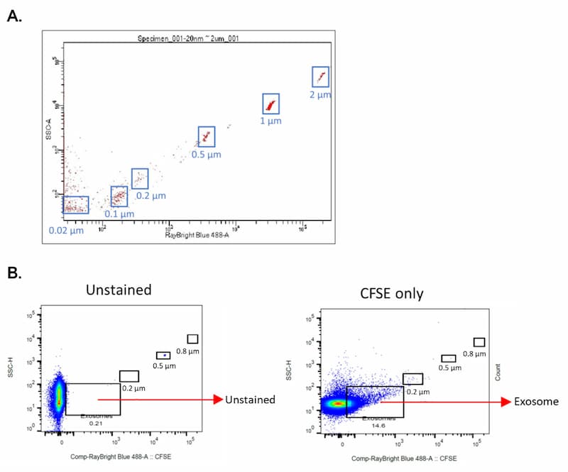
    Purified exosomes are fluorescently labeled with CFSE for sensitive detection by flow cytometry. Labeled samples are analyzed alongside reference beads of known concentration, enabling precise particle counting and normalization across samples. This bead-based approach improves accuracy compared to relative or signal-only measurements and supports cross-experiment comparability.
  • Quality Assessment and Data Confidence
    Flow cytometry analysis provides insight into exosome integrity, labeling efficiency, and sample consistency. By combining CFSE labeling with internal bead standards, we help ensure that exosome measurements reflect true particle counts rather than instrument variability or signal drift.
Exosome Counting and Quality Assessment Image

Exosome identification by flow cytometry. A) Fluorescent reference beads sized from 0.02 µm to 2 µm are used to define the exosome size region based on side scatter and fluorescence signal. B) CFSE labeling enables clear discrimination of exosomes from background debris, revealing a distinct CFSE-positive population at <0.2 µm corresponding to exosomes.

Exosome Phenotyping

We provide flexible exosome phenotyping services, including traditional surface marker profiling and bead-based staining and analysis. These workflows are designed to support a broad range of applications, from basic characterization to functional and disease-focused studies.

  • Surface Marker Profiling
    Exosome surface markers provide critical insight into origin, identity, and biological function. Our surface marker profiling services enable researchers to assess exosome quality, compare populations, and evaluate biologically relevant phenotypes. Researchers may select specific markers of interest or collaborate with our team to design a customized panel aligned with defined research goals.
  • Bead-Based Exosome Staining & Analysis
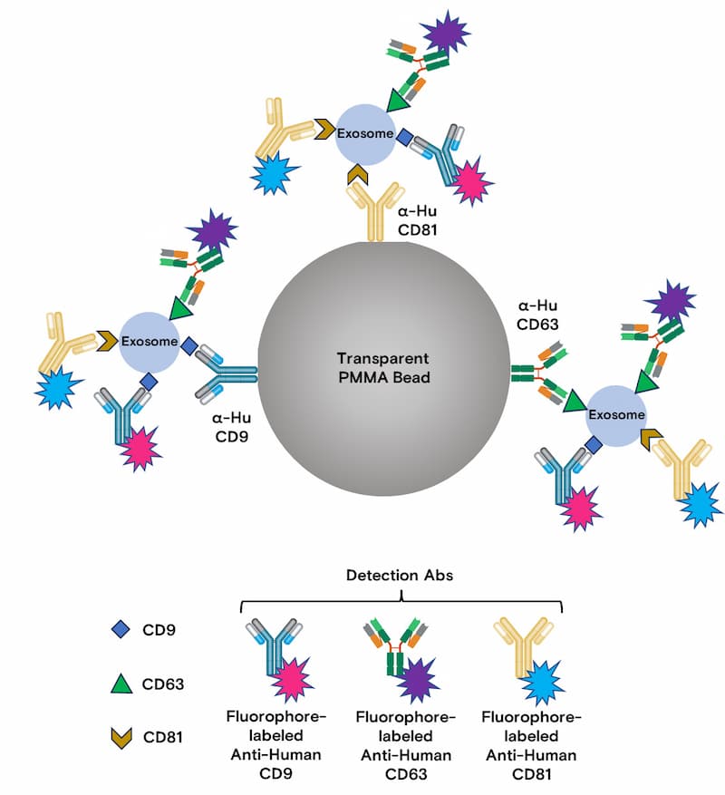
    Bead-based staining enables high-confidence identification and comparative analysis of exosome surface markers by coupling vesicles to antibody-coated beads for flow cytometric detection.

    Workflow:
    1. PMMA beads conjugated with CD9, CD63, and CD81 antibodies*
    2. Incubation with exosome samples
    3. Fluorescent staining with antibodies against CD9, CD63, and CD81
    4. Flow cytometric analysis with isotype controls

Exosome Phenotyping Image 1

*CD9, CD63, and CD81 are commonly expressed exosome markers. Panels can be customized to target additional or alternative markers based on specific research needs.

Exosome Phenotyping Image 2


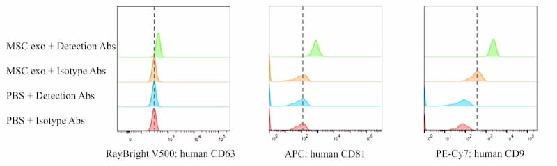
Bead-based phenotyping of MSC-derived exosomes by flow cytometry.
MSC-derived exosomes were captured on antibody-coated PMMA Beads and analyzed for canonical surface markers CD63, CD81, and CD9. Following incubation with exosome samples (PBS used as a negative control), captured vesicles were detected using a mixture of fluorophore-conjugated detection antibodies, with matched isotype controls to confirm specificity. By coupling exosomes to beads, this approach amplifies signal strength and enables clear, side-by-side comparison of marker expression, delivering confident and reproducible exosome phenotyping by flow cytometry.

Exosome Production

RayBiotech offers comprehensive exosome production services, including cell culture and expansion as well as exosome isolation and purification. Post-purification exosome counting and quality assessment can also be performed using the methods described above to ensure consistency and reproducibility.

Cell Culture Platforms
We employ optimized culture conditions using exosome-depleted media to ensure product purity and reproducibility.

Small-Scale Culture

  • Controlled cell expansion and conditioned media harvesting

Large-Scale Bioreactor Production

  • Stirred-Tank Bioreactors
    • Ideal for suspension cell cultures
    • Supports high cell density with real-time monitoring
  • Stirred-Tank Bioreactors with Microcarriers
    • Optimized for adherent cell cultures
    • High yield per volume
    • Preferred for MSC- and stem cell-derived therapeutic exosomes

Isolation and Purification Platforms
RayBiotech utilizes hybrid platforms that integrate multiple isolation and purification strategies to maximize yield, purity, and scalability.

Polymer Precipitation–SEC Workflow:

PEG precipitation → SEC → sterile filtration

  • High yield (60–85%)
  • High purity
  • Scalable and continuous processing

Polymer Precipitation–Affinity Workflow:

PEG precipitation → antibody/ligand capture → elution → sterile filtration

  • Selective purification of specific exosome subpopulations
  • High specificity with moderate yield
  • Suitable for targeted and engineered exosomes

Discuss your project requirements

Request a Quote
Products
  • Multiplex Assays
  • ELISA Kits
  • Proteins and Peptides
  • Antibodies
  • Flow Cytometry
  • Assay Kits
  • Molecular Biology

Sign up to get promotions on your favorite research tools and research updates.

Sign up to Newsletter (opens in new tab)
Services
  • Multiplex Assay Services
  • ELISA Services
  • Antibody Services
  • Custom Protein Services
  • CRO Services
  • Flow Cytometry Services
  • Simoa – Single Molecule Array Services
Resources
  • Manual Protocols
  • Resource Library
  • Learning Center
  • Array Analysis Tools
  • Publications / Citations
  • Publications by RayBiotech Scientists
About Us
  • About RayBiotech
  • Careers
  • Quality Systems
  • Business Partnerships
  • News & Events
  • Reducing Our Emissions
Contact Us
  • Contacts
  • Distributors
  • Certified Service Providers
ISO 13485:2016cGMP
ISO 17025:2017CLIA
©2007-2026 RayBiotech, Inc. All rights reserved.Life Science Web Design By Supreme
  • Privacy Policy
  • Terms & Conditions
  • ISO Certification
  • Risk Free Guarantee
  • Promotions

Your cart is empty.