The sandwich ELISA antibody pair detects Human IgG. Will not detect Fc tagged proteins/peptides.
Compatible Sample Types
Plasma, Serum
Recommended Dilution (Serum/Plasma)
~10,000,000 fold
Quantitative/Semi-Quantitative
Quantitative
Solid Support
96-well Microplate
Design Principle
Sandwich-based
Method Of Detection
Colorimetric
Shipping Type
Blue ice
Storage
≤-20°C
Target Background
Immunoglobulin G (IgG) is an antibody that protects the body from infections, it can bind to viruses, bacteria and fungi. It is the most abundant antibody found in blood, lymph fluid, cerebrospinal fluid and peritoneal fluid and is essential in the humoral immune response. IgG uses agglutination to immobilize pathogens and allows phagocytic immune cells to recognize the pathogen by using opsonization. It can do this because it is a monomeric immunoglobulin made of two heavy chains and two light chains, making up two antigen binding sites. IgG is highly variable due to its composition of amino acids in the terminal portion. The C terminal is used for binding with the cell membrane, the complement system and allows it to pass through the placenta, thus providing protection to the fetus, which no other immunoglobulin can do. Thanks to the abundance and specificity towards antigens, IgG is the principal antibody used in immunological research and clinical diagnostics, making it a great option for ELISA. IgG is retained in the body for long periods of time, the longevity in serum makes it useful for immunization. Detection of IgG is used for indicating prior infection and vaccination.
Product Features
Strip plates and additional reagents allow for use in multiple experiments
Quantitative protein detection
Establishes normal range
The best products for confirmation of antibody array data
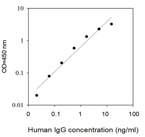
Standard Curves
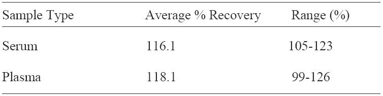
Spiking & Recovery Results
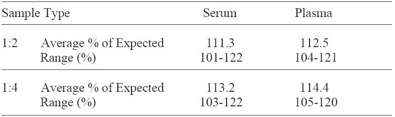
Linearity Results
Typical Data
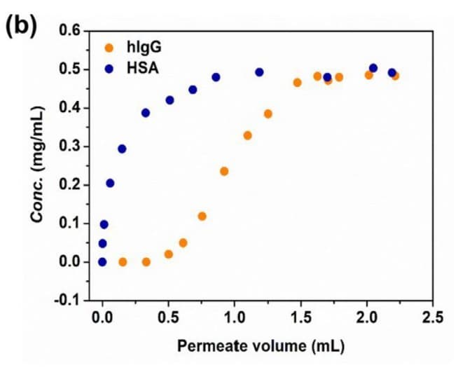
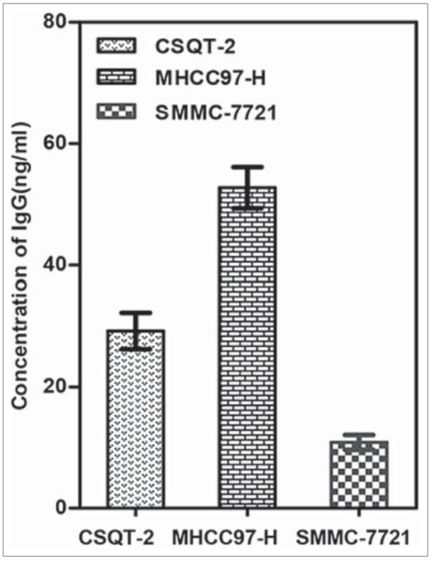
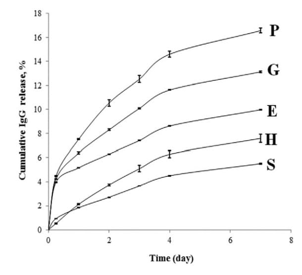
ELH-IGG was used to quantify total plasma IgG levels in rhabdomyosarcoma patient samples to normalize anti-FGF8 autoantibody measurements at diagnosis. Figure adapted from Poli et al., OncoImmunology, 2022. ELH-IGG was used to quantify human IgG recovery from diluted human serum following purification with polyVIM material. Figure adapted from Lei et al., Journal of Chromatography A, 2020. ELH-IGG was used to quantify IgG concentrations in supernatants of hepatocellular carcinoma cell lines, demonstrating that HCC cells secrete IgG with higher levels observed in highly metastatic lines. Figure adapted from Yi et al., OncoImmunology, 2015. ELH-IGG was used to measure biologically active IgG released over 7 days from poly(ε-caprolactone) microspheres formulated with different excipients. Figure adapted from Erdemli et al., Materials Science and Engineering C, 2015.
Kit Components
Pre-Coated 96-well Strip Microplate
Wash Buffer
Stop Solution
Assay Diluent(s)
Lyophilized Standard
Biotinylated Detection Antibody
Streptavidin-Conjugated HRP
TMB One-Step Substrate
Additional Materials Required
Distilled or deionized water
Precision pipettes to deliver 2 µL to 1 µL volumes
Adjustable 1-25 µL pipettes for reagent preparation
100 µL and 1 liter graduated cylinders
Tubes to prepare standard and sample dilutions
Absorbent paper
Microplate reader capable of measuring absorbance at 450nm
Log-log graph paper or computer and software for ELISA data analysis
Assay Procedure Summary
Prepare all reagents, samples and standards as instructed in the manual.
Add 100 µL of standard or sample to each well.
Incubate 2.5 h at RT or O/N at 4°C.
Add 100 µL of prepared biotin antibody to each well.
Incubate 1 h at RT.
Add 100 µL of prepared Streptavidin solution to each well.
Incubate 45 min at RT.
Add 100 µL of TMB One-Step Substrate Reagent to each well.
Incubate 30 min at RT.
Add 50 µL of Stop Solution to each well.
Read at 450 nm immediately.
Storage/Stability
The entire kit may be stored at -20°C for up to 1 year from the date of shipment. Avoid repeated freeze-thaw cycles. The kit may be stored at 4°C for up to 6 months. For extended storage, it is recommended to store at -80°C.