The antibody pair provided in this kit recognizes human H2AX phosphorylated at Serine-139 and total H2AX.
Compatible Sample Types
Tissue Lysates, Cell Lysates
Accession Number
P16104
Gene Id
3014
Gene Symbols
H2AFX, H2AX
Protein Name / Synonyms
Histone H2AX (H2a/x) (Histone H2A.X)
Quantitative/Semi-Quantitative
Semi-Quantitative
Solid Support
96-well Microplate
Design Principle
Sandwich-based
Method Of Detection
Colorimetric
Research Area
Post-Translational Modifications, Phosphorylation, DNA Damage
Shipping Type
Blue ice
Storage
-20°C
Introduction
RayBio® Phospho-H2AX (S139) and Total H2AX ELISA kit is a very rapid, convenient and sensitive assay kit that can monitor the activation or function of important biological pathways in human cell lysates. By determining phosphorylated H2AX protein in your experimental model system, you can verify pathway activation in your cell lysates. You can simultaneously measure numerous different cell lysates without spending excess time and effort in performing a Western Blotting analysis.
This Sandwich ELISA kit is an in vitro enzyme-linked immunosorbent assay for the measurement of human phospho-H2AX and total H2AX. An anti-pan H2AX antibody has been coated onto a 96-well plate. Samples are pipetted into the wells and H2AX present in a sample is bound to the wells by the immobilized antibody. The wells are washed and rabbit anti-H2AX (S139) antibody is used to detect phosphorylated H2AX or rabbit anti-H2AX antibody is used to detect pan H2AX. After washing away unbound antibody, HRP-conjugated anti-rabbit IgG is pipetted to the wells. The wells are again washed, a TMB substrate solution is added to the wells and color develops in proportion to the amount of H2AX (S139) or pan H2AX bound. The Stop Solution changes the color from blue to yellow, and the intensity of the color is measured at 450 nm.
Product Features
Simultaneously measure phosphorylated protein and pan protein in one experiment (for normalization purpose)
Screen numerous different cell lysates without performing a Western Blot analysis
Minimal hands-on time, convenient, and non-radioactive material
Kit Components
Pre-Coated 96-well Strip Microplate
Wash Buffer
Anti-Phospho Antibody
Anti-Pan Antibody (*not included in phospho-only version)
HRP-Conjugated Secondary Antibody
Assay Diluent
TMB One-Step Substrate
Stop Solution
Lysis Buffer
Positive Control Sample
Additional Materials Required
Distilled or deionized water
100 ml and 1 liter graduated cylinders
Tubes to prepare sample dilutions
Protease and Phosphatase inhibitors
Precision pipettes to deliver 2 µl to 1 ml volumes
Adjustable 1-25 ml pipettes for reagent preparation
Benchtop rocker or shaker
Microplate reader capable of measuring absorbance at 450 nm
Assay Procedure Summary
Prepare all reagents and samples as instructed in the manual.
Add 100 µl of sample or positive control to each well.
Incubate 2.5 h at RT or O/N at 4 °C.
Add 100 µl of prepared primary antibody to each well.
Incubate 1 h at RT.
Add 100 µl of prepared 1X HRP-Streptavidin to each well.
Incubate 1 h at RT.
Add 100 µl of TMB One-Step Substrate Reagent to each well.
Incubate 30 min at RT.
Add 50 µl of Stop Solution to each well.
Read at 450 nm immediately.
Typical Data
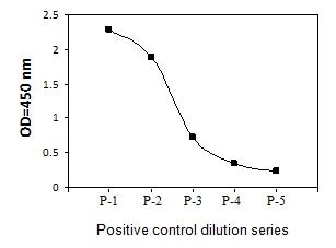
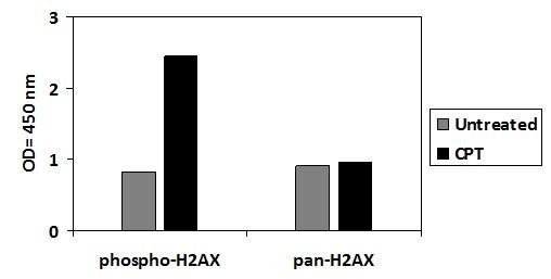
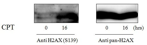
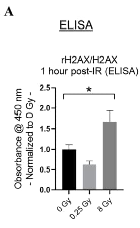
Jurkat cells were treated with CPT at 37°C for 16 hours. Solubilize cells at 4 x 10⁷ cells/ml in Cell Lysate Buffer. Serial dilutions of lysates were analyzed in this ELISA. Please see step 3 of Part VI Reagent Preparation for detail. Jurkat cells were treated or untreated with CPT for 16 hours. Cell lysates were analyzed using this phosphoELISA and Western Blot. RayBio® Phospho-H2AX (Ser139) and Total H2AX ELISA Kits were used to assess radiation-induced DNA damage in human microvascular endothelial cells. Figure adapted from Wang et al., Cells, 2024.
Storage/Stability
Upon receipt, the kit should be stored at €“20°C. Please use within 6 months from the date of shipment. After initial use, Wash Buffer Concentrate (Item B), Assay Diluent (Item E), TMB One-Step Substrate Reagent (Item H), Stop Solution (Item I) and Cell Lysate Buffer (Item J) should be stored at 4°C to avoid repeated freeze-thaw cycles. Return unused wells to the pouch containing desiccant pack, reseal along entire edge, and store at €“20°C. Item D, store at 2-8°C for up to one month (store at -20°C for up to 6 months, avoid repeated freeze-thaw cycles). Reconstituted Positive Control (Item K) should be stored at -70°C.
I analyzed EV (extracellular vesicle) samples.
I detected Phospho-H2AX (S139) on my samples with high confidence.
Positive control perfectly work as I expected.
Review by Kang, from OHSU, OR, USA, on Jun 7, 2023