Detects 18 phosphorylated DNA damage response proteins in mouse samples. Suitable for all liquid sample types but intended for use with cell and tissue lysates.
Loading...
Loading...
Loading...
Loading...
| Size | 2 Sample Kit, 4 Sample Kit, 8 Sample Kit |
| Estimated Lead Time | 1-2 business days |
| Species | Mouse |
| Compatible Sample Types | Tissue Lysates, Cell Lysates |
| Quantitative/Semi-Quantitative | Semi-Quantitative |
| Number of Targets Detected | 18 |
| Recommended Applications | Multiplexed Protein Detection; Detection of Relative Protein Expression; Detecting Patterns of Cytokine Expression; Biomarker/ Key Factor Screening; Identifying Key Factors; Confirming a Biological Process |
| Solid Support | Membrane |
| Design Principle | Sandwich-based |
| Method Of Detection | Chemiluminescence |
| Research Area | Post-Translational Modifications, Phosphorylation, DNA Damage |
| Shipping Type | Blue ice |
| Storage | -20°C |
Acetyl Histone H3 (K9), ATM (S1981), ATR (T1989), C-ABL (Y245), CDC2/CDK1 (Y15), CDC25a (S293), CHK1 (S296), CHK2 (T68), Cyclin B1 (S126), H2AX (S139), NBS1 (S343), P53 (S15), Cleaved PARP (D214/D215), PKMYT1 (T495), PLK1 (T210), RB1 (S811), RSK1 (S380), RSK2 (S386)
Your cart is empty.
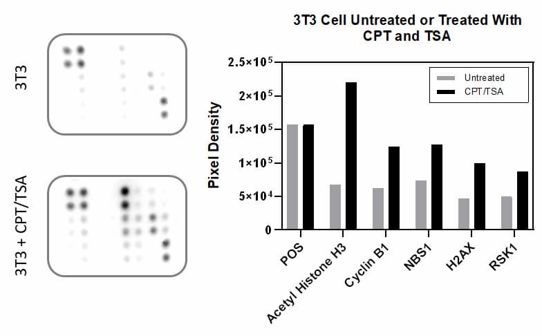
NIH 3T3 cells were either untreated or treated. Data shown are from a 20 second exposure using a chemiluminescence imaging system.
NIH 3T3 cells were either untreated or treated. Data shown are from a 20 second exposure using a chemiluminescence imaging system.