Introduction
Cellular uptake of iron occurs via receptor mediated endocytosis of ligand occupied transferrin receptor into specialized endosomes. Endosomal acidification leads to iron release. The apotransferrin receptor complex is then recycled to the cell surface with a return to neutral pH and the concomitant loss of affinity of apotransferrin for its receptor. Transferrin receptor is necessary for development of erythrocytes and the nervous system (By similarity). Useful in studies of dividing haematopoietic and tumour cell populations, and metabolic activity. A second ligand, the heditary hemochromatosis protein HFE, competes for binding with transferrin for an overlapping C terminal binding site. The antigen is present on most dividing cells, including normally cycling in vivo hematopoietic progenitor cells, mitogenically stimulated cells in vitro, some primary tumor cells and most proliferating cells in vitro.The transferrin receptor has been structurally characterized as a sulfide bound dimer of identical glycoprotein subunits of 95 kDa. The transferrin receptor is not present on resting blood lymphocytes. On PBL, the receptor appears after activation. The expression of transferrin receptor is coordinately regulated with cell growth. Present on T and B cell lines. The soluble (or serum) transferrin receptor (sTfR) is a circulating truncated form of the membrane receptor protein; it is an 85 kDa glycoprotein forming in serum a 320 kDa complex with diferric transferrin.The most important clinical use of the sTfR determination is in the differential diagnosis between iron deficiency anaemia and the anaemia of chronic disease. This antibody is an indicator of proliferation activity. It also has prognostic significance when typing tumors, such as leukemias and lymphomas.
Specificity
Other Names
TFRC; Transferrin receptor protein 1; T9; p90; CD_antigen=CD71; Transferrin receptor protein 1, serum form
NCBI Accession #
NP_001121620.1;NP_003225.2
Format
Type
Purified Rabbit Polyclonal Antibody (Pab)
Calculated Molecular Weight (Da)
84871
Recommended Dilutions
WB: 1:1000; IHC: 1:10-50; FC: 1:10-50
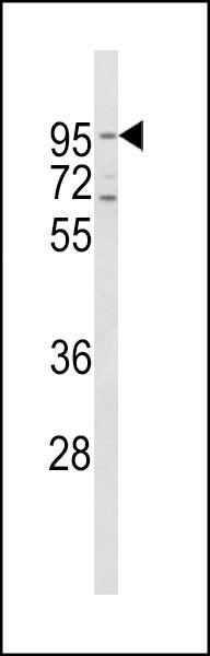 | Western blot analysis of CD71 Antibody (C-term) (Cat# 102-15250) in Hela cell line lysates (35ug/lane). CD71 (arrow) was detected using the purified Pab. |
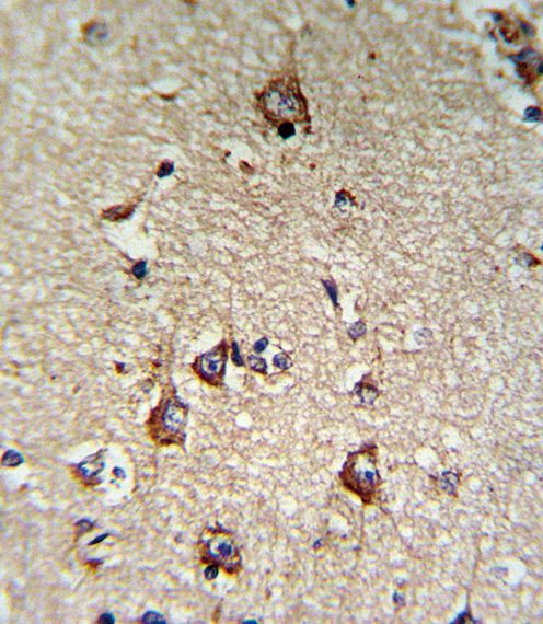 | Formalin-fixed and paraffin-embedded human brain tissue reacted with CD71 Antibody (C-term), which was peroxidase-conjugated to the secondary antibody, followed by DAB staining. This data demonstrates the use of this antibody for immunohistochemistry; clinical relevance has not been evaluated. |
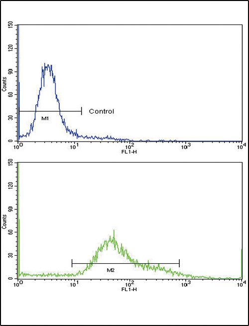 | Flow cytometric analysis of hela cells using CD71 Antibody (C-term)(bottom histogram) compared to a negative control cell (top histogram)FITC-conjugated goat-anti-rabbit secondary antibodies were used for the analysis. |
Antigen Source
HUMAN
Storage/Stability
2-8°C (short-term); -20°C (long-term)
Expiration:
12 months from the date of shipment when stored properly.