Rabbit Anti-Human HDAC9 (N-term) Antibody, 400 ul
Loading...
Loading...
Loading...
Loading...
| Size | 400 µL |
| Estimated Lead Time | 1-2 weeks |
| Species | Human |
| Host Species | Rabbit |
| Accession Number | Q9UKV0 |
| Gene Id | 9734 |
| Gene Symbols | HDAC9 |
| Protein Name / Synonyms | HDAC9 (N-term) |
| Clonality | Polyclonal |
| Preparation | This HDAC9 antibody is generated from rabbits immunized with a KLH conjugated synthetic peptide between 2-32 amino acids from the N-terminal region of human HDAC9. |
| Concentration (lot specific) | 2.0 mg/ml |
| Recommended Applications | Western Blotting, IHC-P, IF, IP |
| Research Area | Angiogenesis, Cardiovascular Disease, Inflammation |
| Shipping Type | Blue ice |
| Storage | -20°C |
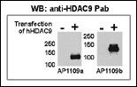 | Both anti-HDAC9 N-term (102-10933) and C-term (102-10934) Pab were tested by WB and IP-WB using HeLa and HeLa-HDAC9 transfected cells. Top figure shows both Pab specifically detect HDAC9 in HeLa-HDAC9 transfected cell but not HeLa alone. |
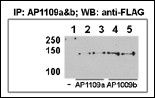 | This figure shows that both Pab can immunoprecipitate (IP) HDAC9 from HeLa-HDAC9 tranfected cells. (Data kindly provided by Dr. Zhigang Yuan, H. Lee Moffitt Cancer Center and Research Institute, Tampa, FL). |
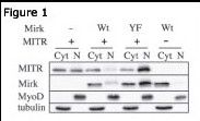 | Figure 1: Immunoblots for MITR (102-10933 HDAC9 N-term antibody), Mirk, MyoD and tubulin proteins are shown for cytoplasmic (Cyt) and nuclear (N) extracts from undifferentiated C2C12 myoblasts. Before cell collection for fractionation, the cells are transfected with plasmids coding for Mirk (Wt), kinase-inactive Mirk (YF) or MITR. Data courtesy of laboratory of Dr. Eileen Friedman. Dept of Pathology, Upstate Medical University, State University of New York. |
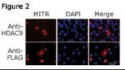 | Figure 2: Immunofluorescence staining of MITR for a compartmentalization study in undifferentiated C2C12 myoblasts transfected with a MITR-expressing plasmid. MITR is detected by using the HDAC9 N-term antibody (top panel) or a FLAG antibody (bottom panel) detecting a FLAG epitope fused at the N-term end of the MITR construct. Data courtesy of laboratory of Dr. Eileen Friedman. Dept of Pathology, Upstate Medical University, State University of New York. |
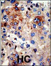 | Formalin-fixed and paraffin-embedded human cancer tissue reacted with the primary antibody, which was peroxidase-conjugated to the secondary antibody, followed by AEC staining. This data demonstrates the use of this antibody for immunohistochemistry; clinical relevance has not been evaluated. BC = breast carcinoma; HC = hepatocarcinoma. |
Your cart is empty.
Anti-HDAC9 (N-term) Antibody
Anti-HDAC9 (N-term) Antibody