Rabbit Anti-Human PI3KC3 (S34) Antibody, 400 ul
Loading...
Loading...
Loading...
Loading...
Anti-PI3KC3 (S34) Antibody
| Size | 400 µL |
| Estimated Lead Time | 1-2 weeks |
| Species | Human |
| Host Species | Rabbit |
| Accession Number | Q8NEB9 |
| Gene Id | 5289 |
| Gene Symbols | PIK3C3 |
| Protein Name / Synonyms | PI3KC3 (S34) |
| Clonality | Polyclonal |
| Preparation | This PI3KC3 antibody is generated from rabbits immunized with a KLH conjugated synthetic peptide between 14-39 amino acids from human PI3KC3. |
| Concentration (lot specific) | 0.5 mg/ml |
| Recommended Applications | Western Blotting, IHC-P, IF |
| Research Area | Autophagy |
| Shipping Type | Blue ice |
| Storage | -20°C |
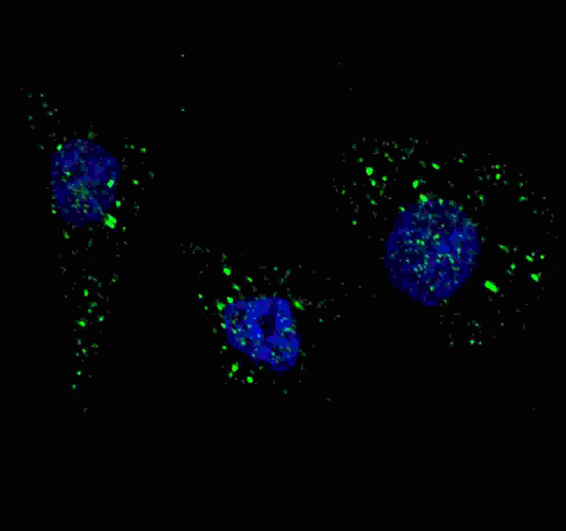 | Fluorescent image of U251 cells stained with PI3KC3 (S34) antibody. U251 cells were treated with Chloroquine (50 µM,16h), then fixed with 4% PFA (20 min), permeabilized with Triton X-100 (0.2%, 30 min). Cells were then incubated with 102-13887 PI3KC3 (S34) primary antibody (1:200, 2 h at room temperature). For secondary antibody, Alexa Fluor® 488 conjugated donkey anti-rabbit antibody (green) was used (1:1000, 1h). Nuclei were counterstained with Hoechst 33342 (blue) (10 µg/ml, 5 min). PI3KC3 immunoreactivity is localized to autophagic vacuoles in the cytoplasm of U251 cells. |
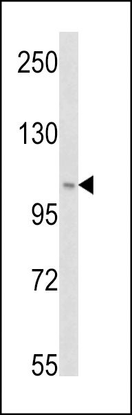 | Western blot analysis of PI3KC3 (S34) (Cat# 102-13887) in Hela cell line lysates (35ug/lane). PI3KC3 (arrow) was detected using the purified Pab. |
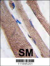 | Formalin-fixed and paraffin-embedded human skeletal muscle tissue reacted with PI3KC3 Antibody (S34), which was peroxidase-conjugated to the secondary antibody, followed by DAB staining. This data demonstrates the use of this antibody for immunohistochemistry; clinical relevance has not been evaluated. |
Your cart is empty.
Anti-PI3KC3 (S34) Antibody