TriMethyl-Histone H3-K27 Polyclonal Antibody
Loading...
Loading...
Loading...
Loading...
| Size | 20 µL, 100 µL |
| Estimated Lead Time | 1-2 weeks |
| Species | Human, Mouse, Rat |
| Host Species | Rabbit |
| Accession Number | Q16695 |
| Gene Id | 8290 |
| Gene Symbols | H3K27ME3 |
| Protein Name / Synonyms | HIST3H3, H3.4, H3/g, H3FT, H3t, histone H3.1t |
| Isotype | IgG |
| Purity | Affinity purification |
| Buffers | PBS with 0.02% sodium azide, 50% glycerol, pH7.3 |
| Recommended Applications | Western Blotting, IHC, IF, IP, ChIP, ChIPseq |
| Shipping Type | Blue ice |
| Storage | -20°C |
Western blot analysis of extracts of various cell lines, using TriMethyl-Histone H3-K27 antibody (144-02363) at 1:1000 dilution.
Secondary antibody: HRP Goat Anti-Rabbit IgG (H+L) at 1:10000 dilution.
Lysates/proteins: 25ug per lane.
Blocking buffer: 3% nonfat dry milk in TBST.
Detection: ECL Basic Kit.
Exposure time: 10s.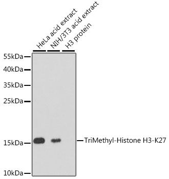
Dot-blot analysis of all sorts of methylation peptides using TriMethyl-Histone H3-K27 antibody (144-02363).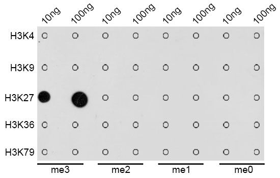
Immunohistochemistry of paraffin-embedded rat ovary using TriMethyl-Histone H3-K27 antibody (144-02363) at dilution of 1:100 (40x lens).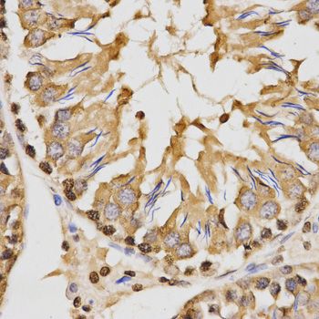
Immunohistochemistry of paraffin-embedded human breast using TriMethyl-Histone H3-K27 antibody (144-02363) at dilution of 1:100 (40x lens).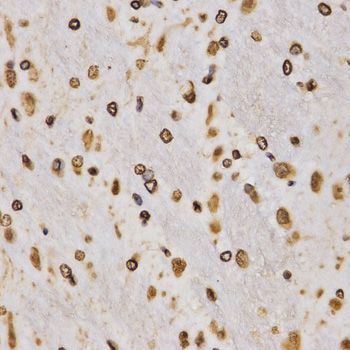
Immunohistochemistry of paraffin-embedded human breast cancer using TriMethyl-Histone H3-K27 antibody (144-02363) at dilution of 1:100 (40x lens).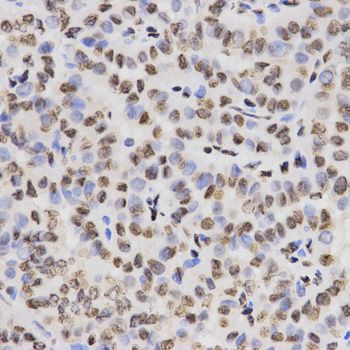
Immunohistochemistry of paraffin-embedded mouse kidney using TriMethyl-Histone H3-K27 antibody (144-02363) at dilution of 1:100 (40x lens).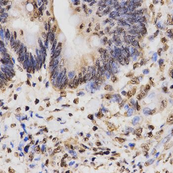
Immunohistochemistry of paraffin-embedded mouse kidney using TriMethyl-Histone H3-K27 antibody (144-02363) at dilution of 1:100 (40x lens).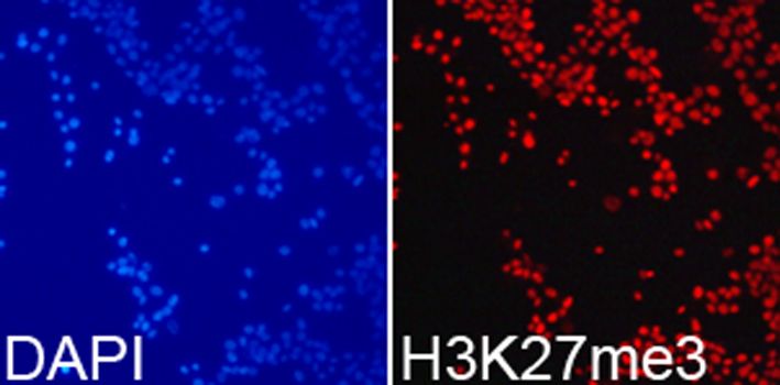
Immunofluorescence analysis of 293T cells using TriMethyl-Histone H3-K27 antibody (144-02363). Blue: DAPI for nuclear staining.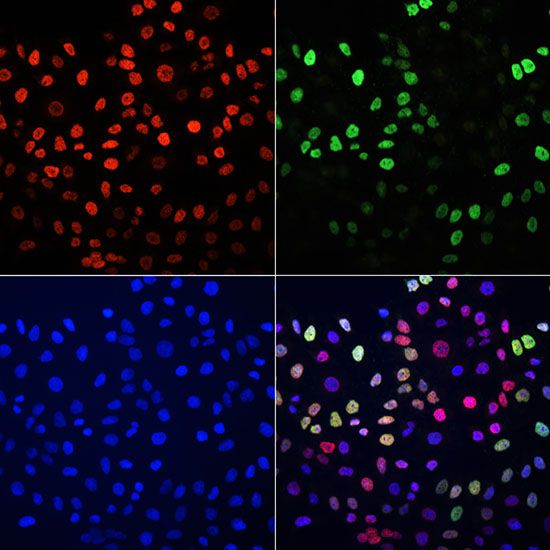
Immunofluorescence analysis of HeLa cells using TriMethyl-Histone H3-K27 antibody(144-02363) and BrdU antibody (Green)(A1482). Blue: DAPI for nuclear staining.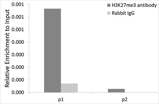
Immunofluorescence analysis of HeLa cells using TriMethyl-Histone H3-K27 antibody (144-02363) at dilution of 1:100. Blue: DAPI for nuclear staining.
Immunofluorescence analysis of C6 cells using TriMethyl-Histone H3-K27 antibody (144-02363) at dilution of 1:100. Blue: DAPI for nuclear staining.
Chromatin immunoprecipitation analysis extracts of 293T cells, using TriMethyl-Histone H3-K27 antibody (144-02363) and rabbit IgG. P1 and P2 were located on ANO2 gene. The amount of immunoprecipitated DNA was checked by quantitative PCR. Histogram was constructed by the ratios of the immunoprecipitated DNA to the input.
Your cart is empty.
Anti-TriMethyl-Histone H3-K27 Antibody
Anti-TriMethyl-Histone H3-K27 Antibody