Biotin-conjugated recombinant human GPC-3 with a C-terminal His-tag, derived from the transfected human HEK293 cells.
Loading...
Loading...
Loading...
Loading...
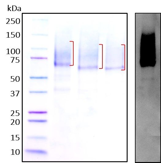
Figure 1. Deglycosylation analysis of purified recombinant proteins. Lane 1, protein ladder (kDa). Lane 2, glycosylated protein (delivered to customers). Lanes 3 and 4, deglycosylated proteins under native and denature conditions respectively.
| Size | 50 µg |
| Estimated Lead Time | 1-2 business days |
| Species | Human |
| Accession Number | P51654-1 |
| Gene Symbols | GPC3 |
| Protein Name / Synonyms | GTR2-2, Intestinal protein OCI-5, MXR7,Glypican-3 alpha subunit, Glypican-3 beta subunit. |
| Expressed Region | Gln25-Asn554 |
| Expression System | HEK293 cells |
| Conjugation | Biotin |
| Tag | C-terminal histidine tag. The primary amino groups (-NH2) at the N-terminus and the side chains of lysine (K) residues were biotin-conjugated using the standard chemical labeling method. |
| Purity | >90% |
| Purity Determined By | SDS-PAGE under reducing conditions and visualized by Coomassie blue staining |
| Molecular Weight (kDa) | Recombinant protein product has a calculated molecular mass of 60 kDa. Due to the abundant glycosylation, it migrates as approximately 65-120 kDa smear protein bands (confrimed by western blotting, right side) in SDS-PAGE under DTT, beta-mercaptoethanol reducing conditions. See deglycosylation analysis image below. |
| Format | Liquid |
| Formulation | Supplied as a 0.2 µm filtered solution in PBS (pH 7.4) |
| Concentration (lot specific) | Lot specific (see the label on the vial), determined by Pierce BCA protein assay |
| Endotoxin Level | <0.5 EU per µg of the protein as determined by the LAL method |
| Shipping Type | Blue ice |
| Storage | -20°C |
Your cart is empty.
Figure 1. Deglycosylation analysis of purified recombinant proteins. Lane 1, protein ladder (kDa). Lane 2, glycosylated protein (delivered to customers). Lanes 3 and 4, deglycosylated proteins under native and denature conditions respectively.