Rabbit Anti-Human ATG5 Antibody, 400 ul
Loading...
Loading...
Loading...
Loading...
| Size | 400 µL |
| Estimated Lead Time | 1-2 weeks |
| Species | Human |
| Host Species | Rabbit |
| Accession Number | Q9H1Y0 |
| Gene Id | 9474 |
| Gene Symbols | ATG5 |
| Protein Name / Synonyms | ATG5 |
| Clonality | Polyclonal |
| Preparation | This ATG5 antibody is generated from rabbits immunized with a KLH conjugated synthetic peptide selected from the full length of human ATG5. |
| Concentration (lot specific) | 1.780 mg/ml |
| Recommended Applications | Western Blotting, IHC-P, IF |
| Research Area | Cardiovascular Disease, Autophagy |
| Shipping Type | Blue ice |
| Storage | -20°C |
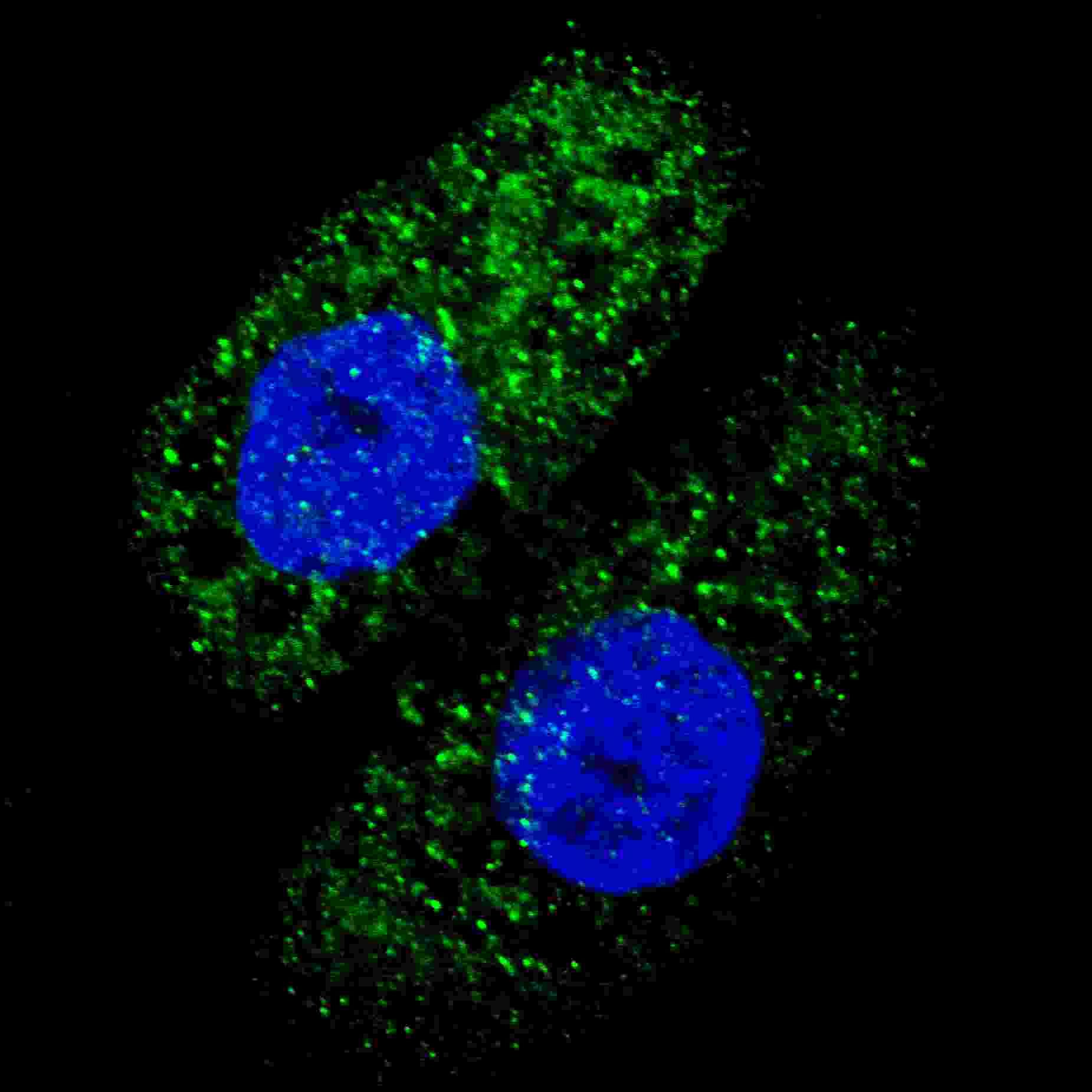 | Fluorescent image of U251 cells stained with ATG5 antibody. U251 cells were treated with Chloroquine (50 µM,16h), then fixed with 4% PFA (20 min), permeabilized with Triton X-100 (0.2%, 30 min). Cells were then incubated with 102-13692 ATG5 primary antibody (1:200, 2 h at room temperature). For secondary antibody, Alexa Fluor® 488 conjugated donkey anti-rabbit antibody (green) was used (1:1000, 1h). Nuclei were counterstained with Hoechst 33342 (blue) (10 µg/ml, 5 min). ATG5 immunoreactivity is localized to autophagic vacuoles in the cytoplasm of U251 cells. |
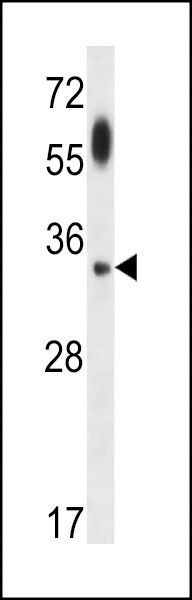 | ATG5 Antibody (Cat# 102-13692) western blot analysis in uterus tumor cell line lysates (35ug/lane).This demonstrates the ATG5 antibody detected the ATG5 protein (arrow). |
| APG5 Antibody (Cat# 102-13692) western blot analysis in uterus tumor cell line lysates (35ug/lane).This demonstrates the APG5 antibody detected the APG5 protein (arrow). |
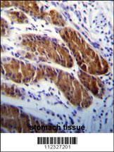 | ATG5 Antibody (Cat# 102-13692)immunohistochemistry analysis in formalin fixed and paraffin embedded human stomach tissue followed by peroxidase conjugation of the secondary antibody and DAB staining.This data demonstrates the use of ATG5 Antibody for immunohistochemistry. Clinical relevance has not been evaluated. |
| APG5 Antibody (Cat# 102-13692b)immunohistochemistry analysis in formalin fixed and paraffin embedded human stomach tissue followed by peroxidase conjugation of the secondary antibody and DAB staining.This data demonstrates the use of APG5 Antibody for immunohistochemistry. Clinical relevance has not been evaluated. |
Your cart is empty.
Anti-ATG5 Antibody
Anti-ATG5 Antibody