Rabbit Anti-Human Connexin 43 (N-term) Antibody, 400 ul
Loading...
Loading...
Loading...
Loading...
| Size | 400 µL |
| Estimated Lead Time | 1-2 weeks |
| Species | Human |
| Host Species | Rabbit |
| Accession Number | P17302 |
| Gene Id | 2697 |
| Gene Symbols | GJA1 |
| Protein Name / Synonyms | Connexin 43 (N-term) |
| Clonality | Polyclonal |
| Preparation | This Connexin 43 antibody is generated from rabbits immunized with a KLH conjugated synthetic peptide between 107-138 amino acids from the N-terminal region of human Connexin 43. |
| Concentration (lot specific) | 2.0 mg/ml |
| Recommended Applications | Western Blotting |
| Research Area | Cardiovascular Disease |
| Shipping Type | Blue ice |
| Storage | -20°C |
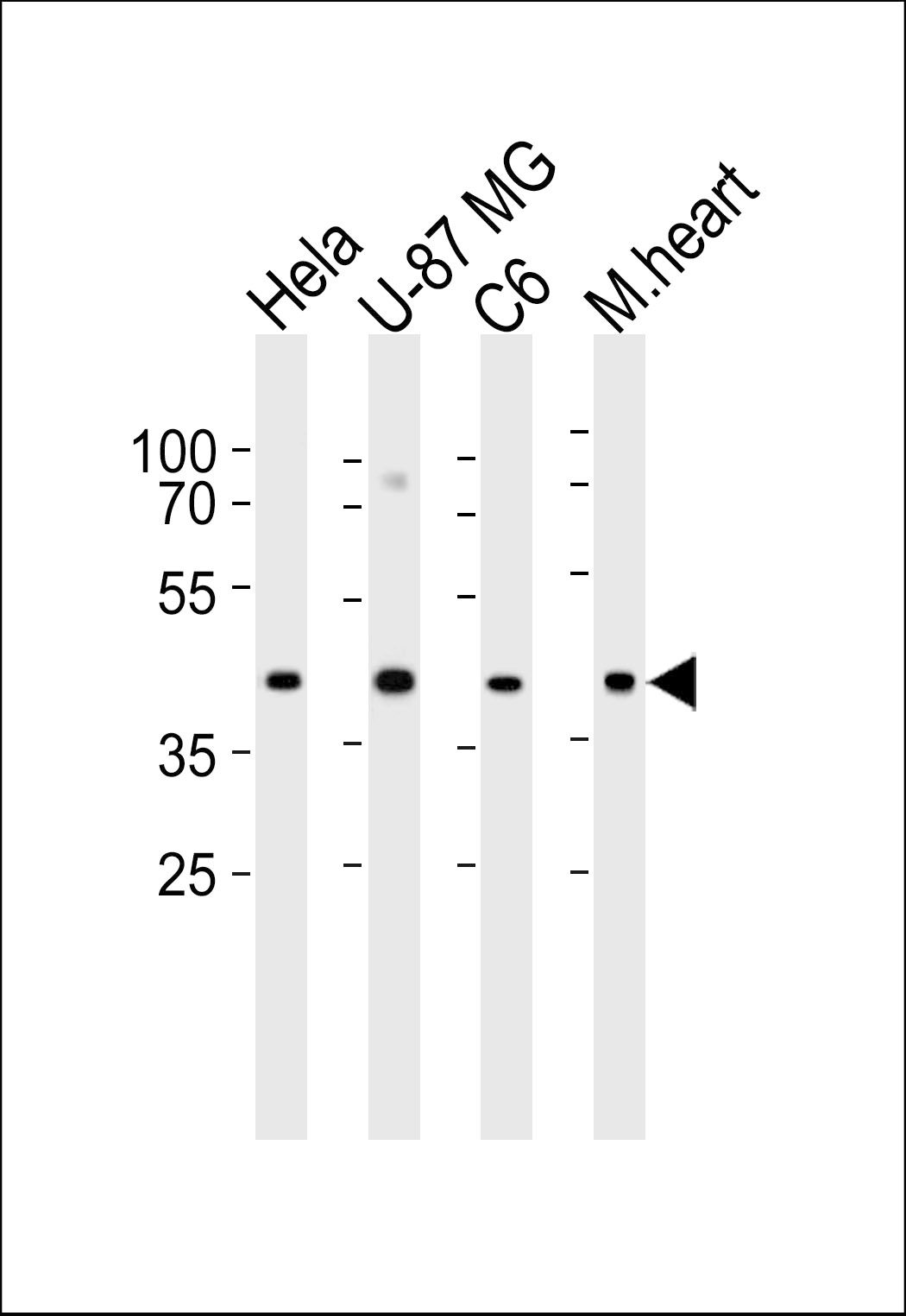 | Western blot analysis of lysates from Hela, U-87 MG, C6 cell line and mouse heart tissue lysate (from left to right), using GJA1 Antibody (N121)(Cat# 102-12784). 102-12784 was diluted at 1:1000 at each lane. A goat anti-rabbit IgG H&L(HRP) at 1:5000 dilution was used as the secondary antibody. Lysates at 35ug per lane. |
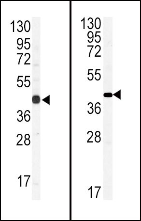 | (LEFT)Western blot analysis of anti-GJA1 Antibody (N-term)(Cat# 102-12784) in CEM cell line lysates (35ug/lane). GJA1(arrow) was detected using the purified Pab.(RIGHT)Western blot analysis of hGJA1-N121.Connexin (Cat# 102-12784) in mouse brain tissue lysates (35ug/lane).GJA1 (arrow) was detected using the purified Pab. |
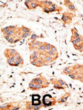 | Formalin-fixed and paraffin-embedded human cancer tissue reacted with the primary antibody, which was peroxidase-conjugated to the secondary antibody, followed by DAB staining. This data demonstrates the use of this antibody for immunohistochemistry; clinical relevance has not been evaluated. BC = breast carcinoma; HC = hepatocarcinoma. |
Your cart is empty.
Anti-Connexin 43 (N-term) Antibody
Anti-Connexin 43 (N-term) Antibody