Lectins are glycan-binding proteins that have been purified from trees, beans, and some fruits. They are highly specific for a given glycan based on their sequence and the different sugar unit structures the glycan contains. The Lectin Array 95 consists of a standard glass slide spotted with 12 identical lectin arrays (1 per well), each containing 95 unique lectins spotted in duplicate, plus positive controls. The slide comes with a 16-well removable gasket which allows for the processing of 12 samples using one slide. Four slides can be nested into a tray, which matches a standard microplate and allows for the automated robotic high-throughput processing of 48 arrays simultaneously. The RayBio® Lectin Array 95 array provides a powerful new tool for glycosylation determination, drug discovery, and biomarker development, all while requiring limited sample volumes.
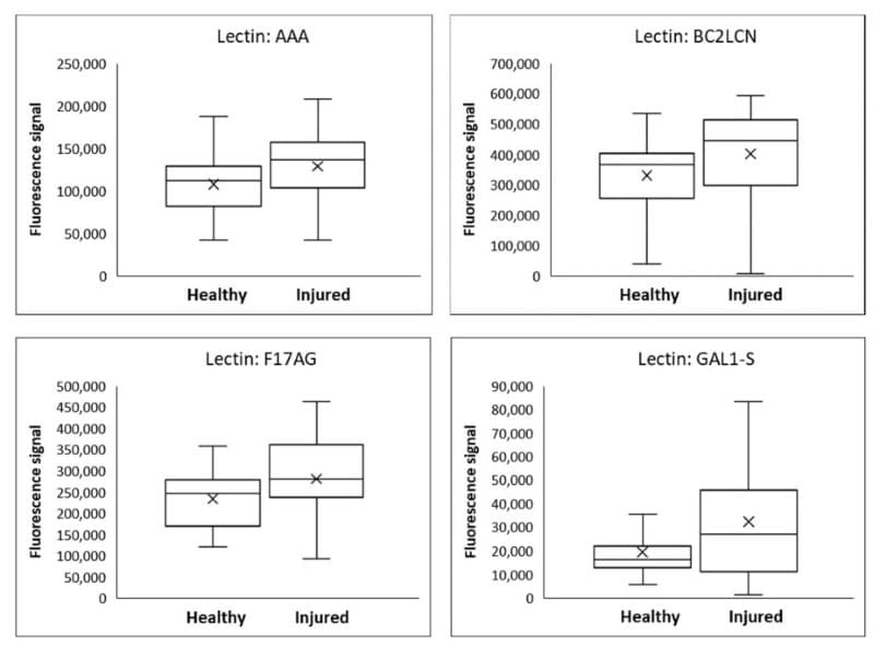
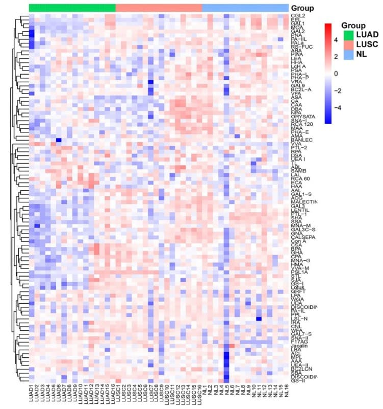
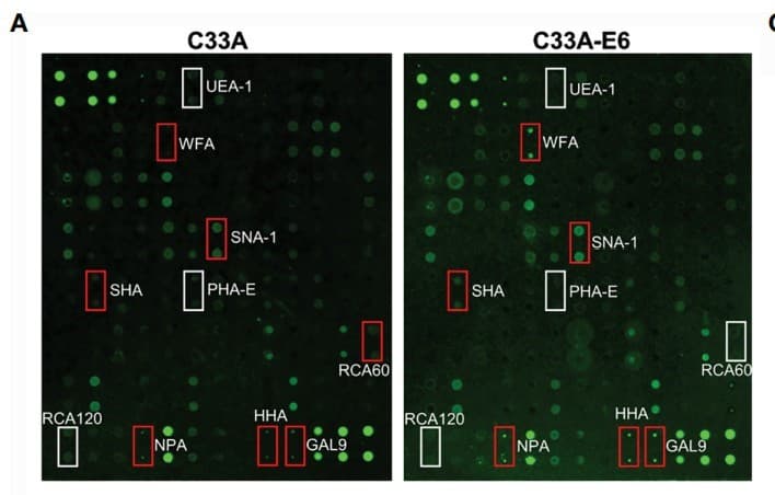
GA-Lectin-95 was used to profile lectin-binding glycan patterns in saliva from pediatric TBI patients and healthy controls, with a few representative lectins shown to illustrate glycan differences associated with brain injury. Figure adapted from Kvist et al., Diagnostics, 2023. GA-Lectin-95 was used to profile plasma glycan-binding patterns in early-stage non-small cell lung cancer patients and healthy controls, with heat map analysis revealing distinct glycosylation signatures associated with disease status. Figure adapted from Li et al., Frontiers in Oncology, 2023. GA-Lectin-95 was used to profile global glycosylation changes in cervical cancer cells expressing HPV16 E6, with representative lectin array images showing increased binding to α2,6-sialylated glycans compared to control cells. Figure adapted from Wang et al., Frontiers in Oncology, 2021.
Application Data/Notes
Application 1 - Detection of Glycans on a Purified Protein
In this application, the RayBio Lectin Array 95 was used to detect specific glycosylations of purified Horseradish Peroxidase (HRP). Lectins BANLEC, BC2L-A, CALSEPA, GNA, HHA, NPA, PA-IIL, and PALa showed strong signals after incubation with 3.3 ug/mL Biotin-HRP followed by detection with streptavidin-fluorescence-dye (Figures A, B and C). The fluorescence signals from BANLEC, BC2L-A, CALSEPA, GNA, HHA, NPA, PA-IIL, and PALa were blocked in a concentration-dependent manner by HRP itself (Figures A and C), indicating that the signals were generated by lectin-HRP binding. These eight lectins are known to exhibit specific binding to mannose, which indicates that HRP contains mannose. After adding increasing amounts of mannose, the signals from BANLEC, BC2L-A, CALSEPA, GNA, HHA, NPA, PA-IIL, and PALa were reduced (Figures A and B).The reduction in signals from increasing concentrations of mannose confirms that HRP protein contains mannose in its glycocalyx. Additionally, the two lectins AAL and RS-FUC (fucose binding specificity) also showed strong interaction with HRP, which indicates the fucosylation of HRP. Overall, the results of the Lectin Array 95 were consistent with published literature regarding HRP glycosylation.
Using the lectin 95 array, we can discover the different glycoprotein profiles of serum samples, cell lysates, or purified glycoproteins. The images above show the profiles of the glycans from different types of samples including human serum, recombinant glycoproteins human HE4 and AFP, mouse TFF2, purified human IgG, and bacterial DE3 cell lysates detected by Biotin labeling and fluorescent dye-streptavidin.
Suggested Applications
Identify and profile the glycans in their samples
Determine whether their biomarker of interest has glycan moieties
Find specific glycan binding ligands in biological samples
Other Applications
Quantitative analysis of lectin-glycoprotein interactions. Example: a concentration series of glycoproteins detected with the lectin array could reveal concentration dependent effects of lectin-glycan binding.
Determine the profile of bacterial cell-surface glycans. Example: Cell lysate from bacteria can be Biotinylated and hybridized to the lectin array. Analysis of the binding pattern and correlation with the known carbohydrate-binding specificities of the lectins can determine the glycans on the cell membrane.
Kit Components
Dialysis Vials
Labeling Reagent
Labeling Buffer
Stop Solution
Lectin Array Glass Slide Assembly
Sample Diluent
20X Wash Buffer I
20X Wash Buffer II
Cy3 equivalent dye-conjugated Streptavidin
Slide Washer/Dryer
Adhesive device sealer
Floating Dialysis Rack
Manual
Additional Materials Required
Detection antibodies of interest (For sandwich-based method only)
Orbital shaker
Laser scanner for fluorescence detection
Aluminum foil
1.5ml Polypropylene microcentrifuge tubes
KCl, NaCl, KH2PO4 and Na2HPO4 (For label-based method only)
Plastic or glass containers, beaker, stir plate and stir bar
Pipettors, pipette tips, ddH2O and other common lab consumables
Assay Procedure Summary
Dry the glass slide
Block array surfaces
Incubate samples (samples need to be biotinylated for the label-based approach)
For the the sandwich-based principle, incubate with a detection antibody cocktail For the label-based principle, incubate the labeled-streptavidin.
Upon receipt, all components of the Raybiotech Lectin Array 95 kit should be stored at -20°C. Once thawed, the glass slide and Cy3 equivalent dye-conjugated Streptavidin should be kept at -20°C and all other components may be stored at 4°C. The entire kit should be used within 6 months of purchase.