Introduction
Recombinant Monkeypox virus (MPXV) proteins derived from bacterial and human cells (HEK293) are spotted in triplicate on a standard glass slide (25 mm x 75 mm x 1 mm), along with positive and negative controls to monitor each incubation step. When liquid sample types, such as serum or purified antibodies, are incubated on the array, "primary" antibodies in the sample will bind to their specific target proteins. Specific primary antibody isotypes (IgG, IgM, or IgA) from serum, plasma, purified antibodies, etc. are then targeted by biotin-conjugated anti-IgG, IgM, or IgA secondary antibodies, respectively, that are specific for the host species.
A fluorophore-conjugated streptavidin molecule is then added, which binds to the biotin on the secondary antibody. The fluorophore enables the detection of the immobilized primary antibody via fluorescence using a laser scanner. The fluorescence signal is proportional to the amount of immobilized antibody. Since each spot represents a unique protein that is known, the specific target(s) bound by the antibodies can be ascertained.
Application Notes
- Vaccine Development: Measure IgG, IgM, or IgA antibody responses to immunization or patients
- Protein-protein Interaction Studies: Determine the interactions between the viral proteins and other molecules, such as proteins, peptides, and small molecules. (Additional reagents and materials not included in the kit may be required.)
Typical Data
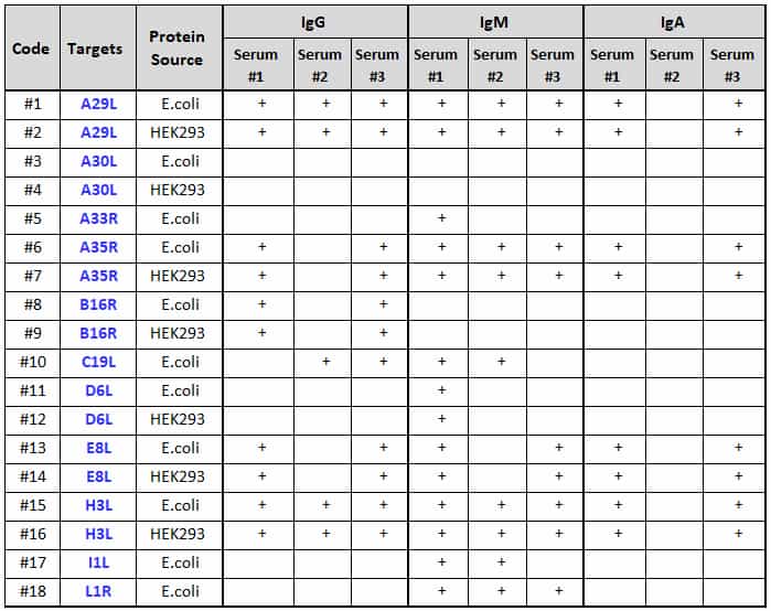
Three serum samples (Serum #1, #2, and #3) from Monkeypox virus infected individuals (June 2022, USA) were assayed using the Raybio® Monkeypox Virus Protein Array G1 (Cat # PAV-MPHG-G1). Three isoforms — IgG, IgM and IgA were simultaneously probed from each sample, revealing a significant immune response to various viral target proteins (Figure 1, Table 1). Interestingly, both bacterial-derived and human cell-derived recombinant viral proteins had similar antibody responses.
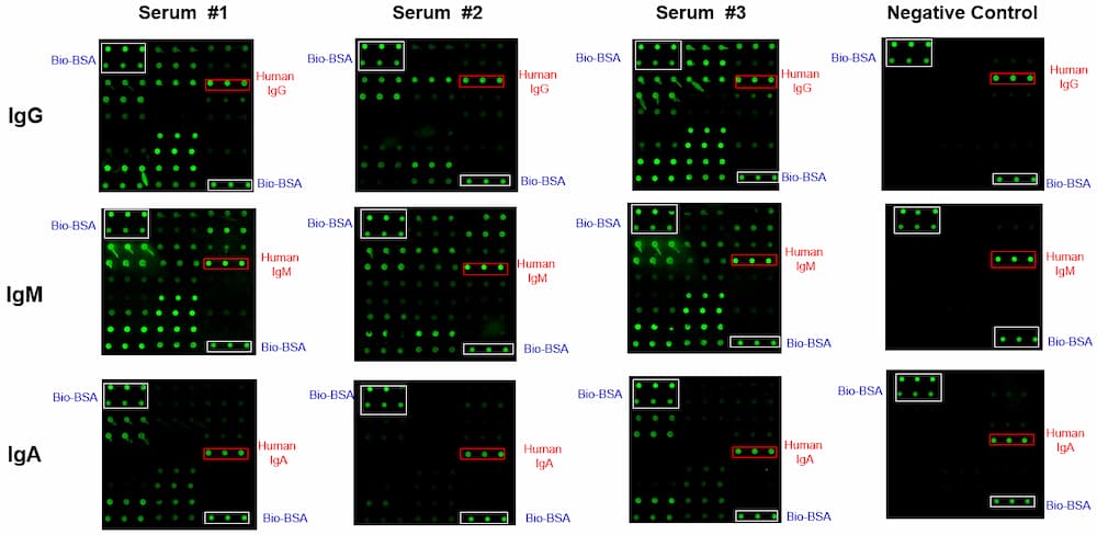
Figure 1. Monkeypox Virus Protein Array G1 run with patient serum samples. Monkeypox Virus Protein Array G1 was printed on a glass-slide array with Bio-BSA and IgG/IgM/IgA positive controls (white and red highlighted boxes, respectively) and 18 E. coli- or HEK293-derived Monkeypox viral proteins (Table 1). Serum samples from three Monkeypox virus infected individuals (Serum #1, #2, and #3) were obtained, diluted 1,000-fold and incubated in the array along with a blocking buffer negative control. The arrays were probed using biotin-conjugated anti-human IgG, IgM, and IgA, respectively. Followed by wash steps, the array was incubated with Cy3 equivalent dye-conjugated streptavidin. The array signals were visualized using a laser scanner capable of measuring signals in the green channel.
Table 1. Immune response of Monkeypox virus infected patients to different MPXV proteins
Product Features
- Small sample volume required
- High density
- High sensitivity
- Large dynamic range
- Suitable for high-throughput assays
Kit Components
- RayBio® Monkeypox virus protein array
- Blocking Buffer
- Wash Buffer I
- Wash Buffer II
- Biotin-conjugated secondary antibody
- Cy3 equivalent dye conjugated streptavidin
- Array accessories (including slide incubation chamber assembly, gasket, adhesive plastic strips, etc.)
Other Materials Required
- Distilled water
- Small plastic boxes or containers
- Orbital shaker or oscillating rocker
- Pipettors, pipette tips and other common lab consumables
- Aluminum foil
- Laser fluorescence scanner
View Compatible Laser Scanners
Don't have a compatible scanner? RayBiotech now offers FREE scanning service for all RayBio glass slide antibody arrays! Learn More
Storage/Stability
Upon receipt, all kit components should be stored at -20 °C to -80 °C until ready to use. Stored properly, the kit will retain complete activity for up to 6 months.