RayBiotech

3607 Parkway Ln Ste 200 Norcross, Georgia 30092

www.RayBiotech.com | info@raybiotech.com

770-729-2992 | +1-888-494-8555

ISO 13485:2016 cGMP

Anti-EGFR/ErbB1 (pSer1045) Antibody

Catalog #: RB-02-0004

Rabbit Anti-Human EGFR/ErbB1 (pSer1045) Antibody

Product Description

Specifications

Size20 µg, 100 µg, 200 µg, 500 µg
Estimated Lead Time1-2 business days
SpeciesHuman
ClonalityPolyclonal
Immunogen
Immunogen was synthetic peptide derived from human EGFR. This antibody was obtained from a rabbit immunized with the immunogen. The IgG fraction of immunized serum was purified by Protein G affinity chromatography.
Immunogen Sequence
SATSNNS(p)TVAC
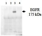
Recommended ApplicationsELISA (recommended work dilution= 1: 5, 000-10, 000), Western Blotting (recommended work dilution= 1-5 µg/ml), (see image) RB-02-0002 (1-5 µg/ml) staining of Hela and A431 lysate. Lane 1 and 2, Hela lysate with and without stimulating, Land 3 and 4, A431 lysate with and without stimulating. Detected by western blot using chemiluminescence.
Reconstitution
Supplied as 100 µg of purified antibody in 10 mM sodium HEPES (pH 7.5),150 mM NaCl, 100 µg/ml BSA and 50% glycerol. Store at –20°C. Do not aliquot the antibody.
Research AreaCancer, HER/ErbB Signaling, Tyrosine Kinase Signaling
Shipping TypeAmbient temperature
Storage-20°C

Description

Description

The epidermal growth factor (EGF) receptor is a 170 kDa transmembrane tyrosine kinase that belongs to the HER/ErbB protein family. Ligand binding results in receptor dimerization, autophosphorylation, activation of downstream signaling and lysosomal degradation . Phosphorylation of EGF receptor (EGFR) at Tyr845 in the kinase domain is implicated in stabilizing the activation loop, maintaining the active state enzyme and providing a binding surface for substrate proteins. c-Src is involved in phosphorylation of EGFR at Tyr845. The SH2 domain of PLC? binds at phospho-Tyr992, resulting in activation of PLC?-mediated downstream signaling. Phosphorylation of EGFR at Tyr1045 creates a major docking site for c-Cbl, an adaptor protein that leads to receptor ubiquitination and degradation following EGFR activation. The GRB2 adaptor protein binds activated EGFR at phospho-Tyr1068. A pair of phosphorylated residues (Tyr1148 and Tyr1173) provides a docking site for the SHC scaffold protein, with both sites involved in MAP kinase signaling activation. Phosphorylation of EGFR at specific serine and threonine residues attenuates EGFR kinase activity. EGFR carboxy-terminal residues Ser1046 and Ser1047 are phosphorylated by CaM kinase II; mutations to either of these serines results in upregulated EGFR tyrosine autokinase activity.

Typical Data

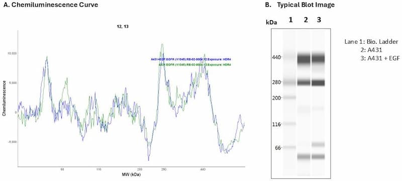
Cell Lysate: A431 + EGF | Secondary Ab: Rabbit | Exposure: HDR | Expected MW (kDa): 134

Specificity

The antibody specifically detects phosphorylated protein derived from A431 cell lysates at various concentrations. In both ELISA and EGFR, this antibody shows no cross-reactivity with other unrelated protein.

Expiration

12 months from the date of shipment when stored properly.