Mouse Anti-Human CD44 Antibody, 400 ul
Loading...
Loading...
Loading...
Loading...
| Size | 400 µL |
| Estimated Lead Time | 1-2 weeks |
| Species | Human |
| Accession Number | P16070 |
| Gene Id | 960 |
| Gene Symbols | CD44 |
| Protein Name / Synonyms | CD44 |
| Clonality | Monoclonal |
| Preparation | This CD44 monoclonal antibody is generated from mouse immunized with CD44 recombinant protein. |
| Concentration (lot specific) | 0.500 mg/ml |
| Recommended Applications | Western Blotting, IHC-P, IF, FC |
| Research Area | Inflammation, Adaptive Immunity |
| Shipping Type | Blue ice |
| Storage | -20°C |
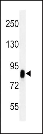 | CD44 antibody (Cat# 102-10131) western blot analysis in Hela cell line lysates (35ug/lane).This demonstrates the CD44 antibody detected the CD44 protein (arrow). |
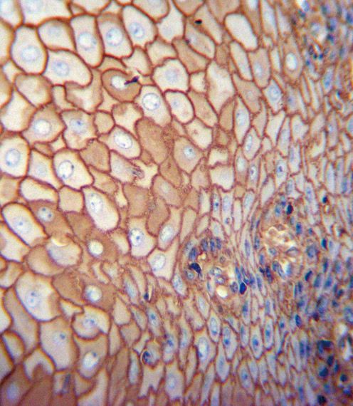 | CD44 antibody (Cat# 102-10131) immunohistochemistry analysis in formalin fixed and paraffin embedded human esophagus carcinoma followed by peroxidase conjugation of the secondary antibody and DAB staining. This data demonstrates the use of the CD44 antibody for immunohistochemistry. Clinical relevance has not been evaluated. |
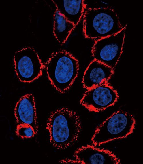 | CD44 antibody (Cat# 102-10131) confocal immunofluorescent analysis with hela cell. 0.01 mg/ml primary antibody was followed by PE-conjugated goat anti-mouse lgG (whole molecule). PE emits red fluorescence. DAPI was used to stain the cell nuclear (blue). |
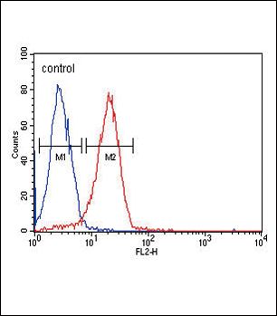 | CD44 Antibody (Cat# 102-10131) flow cytometric analysis of Hela cells (right histogram) compared to a negative control cell (left histogram).PE-conjugated goat-anti-mouse secondary antibodies were used for the analysis. |
Your cart is empty.
Anti-CD44 Antibody
Anti-CD44 Antibody