This product contains purified exosomes isolated from Jurkat cells, an immortalized human CD4+ T-cell line.
Loading...
Loading...
Loading...
Loading...
Jurkat exosomes were captured by bead-based immunoprecipitation, stained with dye-conjugated antibodies against specific surface markers, and analyzed via flow cytometry.
| Size | 50 µg |
| Estimated Lead Time | 1-2 business days |
| Species | Human |
| Format | Purified exosome suspension, frozen |
| Buffers | 0.05 µm (50 nm)-filtered PBS containing 5% trehalose and protease inhibitor cocktail |
| Shipping Type | Dry ice (additional shipping fee) |
| Storage | -80°C |
Exosomes were characterized by flow cytometry to confirm expression of universal exosome markers CD9, CD63, and CD81, as well as additional cell-specific markers. For Jurkat-derived exosomes specifically, CD9, CD63, CD81, CD3, CD4, and CD45 were analyzed. As shown in Figure 1, exosomes showed strong expression of CD9, CD81, and CD3.
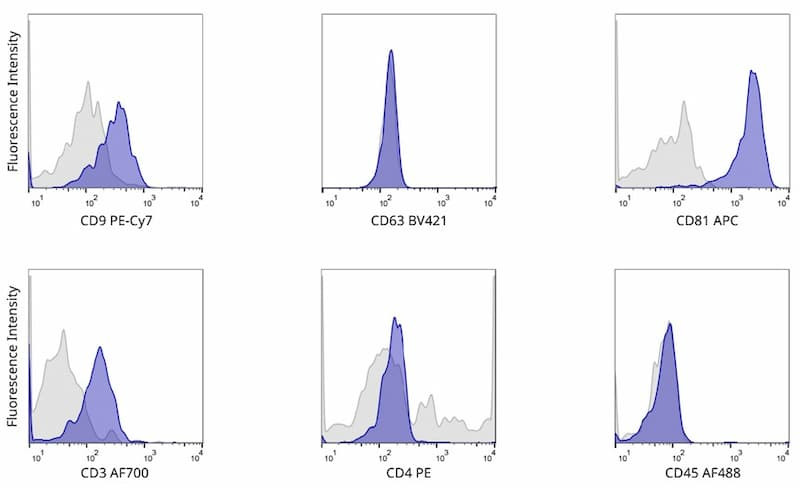
Jurkat exosomes were captured by bead-based immunoprecipitation, stained with dye-conjugated antibodies against specific surface markers, and analyzed via flow cytometry.
Sign up to get promotions on your favorite research tools and research updates.


